Ideal for Aorta Tissue Homogenization
Do you spend lots of time and effort homogenizing aorta tissue samples? The Bullet Blender® tissue homogenizer delivers high quality and superior yields. No other homogenizer comes close to delivering the Bullet Blender’s winning combination of top-quality performance and budget-friendly affordability. See below for an aorta tissue homogenization protocol.
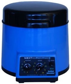
The Bullet Blender®
Save Time, Effort and Get Superior Results
- Consistent and High Yield Results
Run up to 24 samples at the same time under microprocessor-controlled conditions, ensuring experimental reproducibility and high yield. Process samples from 10mg or less up to 3.5g. - No Cross Contamination
No part of the Bullet Blender® ever touches the aorta samples – the sample tubes are kept closed during homogenization. There are no probes to clean between samples. - Samples Stay Cool
Homogenizing causes only a few degrees of heating. Our Gold models keep samples at 4°C - Easy and Convenient to Use
Just place beads and buffer along with your aorta sample in standard tubes, load tubes directly in the Bullet Blender, select time and speed, and press start. - Risk Free Purchase
The Bullet Blender® comes with a 30 day money back guarantee and a 2 year warranty, with a 3 year warranty on the motor. The simple, reliable design enables the Bullet Blenders to sell for a fraction of the price of ultrasonic or other agitation based instruments, yet provides an easier, quicker technique.
Bullet Blender Homogenization Protocol for Aorta Tissue
Sample size |
See the Protocol |
| microcentrifuge tube model (up to 300 mg) | Small aorta tissue samples |
| 5mL tube model (100mg – 1g) | Medium aorta tissue samples |
Selected Publications for Aorta Tissue
See all of our Bullet Blender publications!
Papke, C. L., Tsunezumi, J., Ringuette, L.-J., Nagaoka, H., Terajima, M., Yamashiro, Y., Urquhart, G., Yamauchi, M., Davis, E. C., & Yanagisawa, H. (2015). Loss of fibulin-4 disrupts collagen synthesis and maturation: implications for pathology resulting from EFEMP2 mutations. Human Molecular Genetics, 24(20), 5867–5879. https://doi.org/10.1093/hmg/ddv308
Reho, J. J., Zheng, X., Asico, L. D., & Fisher, S. A. (2015). Redox signaling and splicing dependent change in myosin phosphatase underlie early versus late changes in NO vasodilator reserve in a mouse LPS model of sepsis. American Journal of Physiology - Heart and Circulatory Physiology, 308(9), H1039–H1050. https://doi.org/10.1152/ajpheart.00912.2014
Rotllan, N., Chamorro-Jorganes, A., Araldi, E., Wanschel, A. C., Aryal, B., Aranda, J. F., Goedeke, L., Salerno, A. G., Ramirez, C. M., Sessa, W. C., Suarez, Y., & Fernandez-Hernando, C. (2015). Hematopoietic Akt2 deficiency attenuates the progression of atherosclerosis. The FASEB Journal, 29(2), 597–610. https://doi.org/10.1096/fj.14-262097
Carlström, M., Liu, M., Yang, T., Zollbrecht, C., Huang, L., Peleli, M., Borniquel, S., Kishikawa, H., Hezel, M., Persson, A. E. G., Weitzberg, E., & Lundberg, J. O. (2015). Cross-talk Between Nitrate-Nitrite-NO and NO Synthase Pathways in Control of Vascular NO Homeostasis. Antioxidants & Redox Signaling, 23(4), 295–306. https://doi.org/10.1089/ars.2013.5481
Gao, N., Huang, J., He, W., Zhu, M., Kamm, K. E., & Stull, J. T. (2013). Signaling through Myosin Light Chain Kinase in Smooth Muscles. Journal of Biological Chemistry, 288(11), 7596–7605. https://doi.org/10.1074/jbc.M112.427112
Kuang, S.-Q., Kwartler, C. S., Byanova, K. L., Pham, J., Gong, L., Prakash, S. K., Huang, J., Kamm, K. E., Stull, J. T., Sweeney, H. L., & Milewicz, D. M. (2012). Rare, Nonsynonymous Variant in the Smooth Muscle-Specific Isoform of Myosin Heavy Chain, MYH11, R247C, Alters Force Generation in the Aorta and Phenotype of Smooth Muscle Cells. Circulation Research, 110(11), 1411–1422. https://doi.org/10.1161/CIRCRESAHA.111.261743
Fox, K. A., Longo, M., Tamayo, E., Gamble, P., Makhlouf, M., Mateus, J. F., & Saade, G. R. (2012). Sex-specific effects of nicotine exposure on developmental programming of blood pressure and vascular reactivity in the C57Bl/6J mouse. American Journal of Obstetrics and Gynecology, 207(3), 208.e1-208.e9. https://doi.org/10.1016/j.ajog.2012.06.021
Shai, S.-Y., Sukhanov, S., Higashi, Y., Vaughn, C., Rosen, C. J., & Delafontaine, P. (2011). Low circulating insulin-like growth factor I increases atherosclerosis in ApoE-deficient mice. AJP: Heart and Circulatory Physiology, 300(5), H1898–H1906. https://doi.org/10.1152/ajpheart.01081.2010
Fox, K. A., Longo, M., Tamayo, E., Kechichian, T., Bytautiene, E., Hankins, G. D. V., Saade, G. R., & Costantine, M. M. (2011). Effects of pravastatin on mediators of vascular function in a mouse model of soluble Fms-like tyrosine kinase-1–induced preeclampsia. American Journal of Obstetrics and Gynecology, 205(4), 366.e1-366.e5. https://doi.org/10.1016/j.ajog.2011.06.083
Rayner, K. J., Suarez, Y., Davalos, A., Parathath, S., Fitzgerald, M. L., Tamehiro, N., Fisher, E. A., Moore, K. J., & Fernandez-Hernando, C. (2010). MiR-33 Contributes to the Regulation of Cholesterol Homeostasis. Science, 328(5985), 1570–1573. https://doi.org/10.1126/science.1189862
Lee, Y. W., Lee, W. H., & Kim, P. H. (2010). Role of NADPH oxidase in interleukin-4-induced monocyte chemoattractant protein-1 expression in vascular endothelium. Inflammation Research, 59(9), 755–765. https://doi.org/10.1007/s00011-010-0187-3
Lee, Y. W., Lee, W. H., & Kim, P. H. (2010). Oxidative mechanisms of IL-4-induced IL-6 expression in vascular endothelium. Cytokine, 49(1), 73–79. https://doi.org/10.1016/j.cyto.2009.08.009
Hou, C. J.-Y., Tsai, C.-H., Su, C.-H., Wu, Y.-J., Chen, S.-J., Chiu, J.-J., Shiao, M.-S., & Yeh, H.-I. (2008). Diabetes Reduces Aortic Endothelial Gap Junctions in ApoE-deficient Mice: Simvastatin Exacerbates the Reduction. Journal of Histochemistry and Cytochemistry, 56(8), 745–752. https://doi.org/10.1369/jhc.2008.950816