Ideal for Eye Tissue Homogenization
Do you spend lots of time and effort homogenizing eye tissue samples? The Bullet Blender® tissue homogenizer delivers high quality and superior yields. No other homogenizer comes close to delivering the Bullet Blender’s winning combination of top-quality performance and budget-friendly affordability. See below for an eye tissue homogenization protocol.
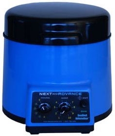
The Bullet Blender®
Save Time, Effort and Get Superior Results
- Consistent and High Yield Results
Run up to 24 samples at the same time under microprocessor-controlled conditions, ensuring experimental reproducibility and high yield. Process samples from 10mg or less up to 3.5g. - No Cross Contamination
No part of the Bullet Blender® ever touches the eye tissue samples – the sample tubes are kept closed during homogenization. There are no probes to clean between samples. - Samples Stay Cool
Homogenizing causes only a few degrees of heating. Our Gold models keep samples at 4°C. - Easy and Convenient to Use
Just place beads and buffer along with your eye tissue sample in standard tubes, load tubes directly in the Bullet Blender, select time and speed, and press start. - Risk Free Purchase
The Bullet Blender® comes with a 30 day money back guarantee and a 2 year warranty, with a 3 year warranty on the motor. The simple, reliable design enables the Bullet Blenders to sell for a fraction of the price of ultrasonic or other agitation based instruments, yet provides an easier, quicker technique.
Bullet Blender Homogenization Protocol for Eye Tissue
Sample size |
See the Protocol |
| microcentrifuge tube model (up to 300mg) | Small eye samples |
| 5mL tube model (100mg – 1g) | Medium eye samples |
| 50mL tube model (100mg – 3.5g) | Large eye samples |
Selected Publications for Eye Tissue
See all of our Bullet Blender publications!
2474232
eye
items
1
0
date
desc
3154
https://www.nextadvance.com/wp-content/plugins/zotpress/
%7B%22status%22%3A%22success%22%2C%22updateneeded%22%3Afalse%2C%22instance%22%3A%22zotpress-4ea993c279b1bd99413ebcbcc4f7f4b1%22%2C%22meta%22%3A%7B%22request_last%22%3A0%2C%22request_next%22%3A0%2C%22used_cache%22%3Atrue%7D%2C%22data%22%3A%5B%7B%22key%22%3A%22UW9VGJ3J%22%2C%22library%22%3A%7B%22id%22%3A2474232%7D%2C%22meta%22%3A%7B%22creatorSummary%22%3A%22Zhou%20et%20al.%22%2C%22parsedDate%22%3A%222016-08%22%2C%22numChildren%22%3A2%7D%2C%22bib%22%3A%22%3Cdiv%20class%3D%5C%22csl-bib-body%5C%22%20style%3D%5C%22line-height%3A%202%3B%20padding-left%3A%201em%3B%20text-indent%3A-1em%3B%5C%22%3E%5Cn%20%20%3Cdiv%20class%3D%5C%22csl-entry%5C%22%3EZhou%2C%20Y.%2C%20Bennett%2C%20T.%20M.%2C%20%26amp%3B%20Shiels%2C%20A.%20%282016%29.%20Lens%20ER-stress%20response%20during%20cataract%20development%20in%20Mip-mutant%20mice.%20%3Ci%3EBiochimica%20et%20Biophysica%20Acta%20%28BBA%29%20-%20Molecular%20Basis%20of%20Disease%3C%5C%2Fi%3E%2C%20%3Ci%3E1862%3C%5C%2Fi%3E%288%29%2C%201433%26%23x2013%3B1442.%20%3Ca%20href%3D%27https%3A%5C%2F%5C%2Fdoi.org%5C%2F10.1016%5C%2Fj.bbadis.2016.05.003%27%3Ehttps%3A%5C%2F%5C%2Fdoi.org%5C%2F10.1016%5C%2Fj.bbadis.2016.05.003%3C%5C%2Fa%3E%3C%5C%2Fdiv%3E%5Cn%3C%5C%2Fdiv%3E%22%2C%22data%22%3A%7B%22itemType%22%3A%22journalArticle%22%2C%22title%22%3A%22Lens%20ER-stress%20response%20during%20cataract%20development%20in%20Mip-mutant%20mice%22%2C%22creators%22%3A%5B%7B%22creatorType%22%3A%22author%22%2C%22firstName%22%3A%22Yuefang%22%2C%22lastName%22%3A%22Zhou%22%7D%2C%7B%22creatorType%22%3A%22author%22%2C%22firstName%22%3A%22Thomas%20M.%22%2C%22lastName%22%3A%22Bennett%22%7D%2C%7B%22creatorType%22%3A%22author%22%2C%22firstName%22%3A%22Alan%22%2C%22lastName%22%3A%22Shiels%22%7D%5D%2C%22abstractNote%22%3A%22Major%20intrinsic%20protein%20%28MIP%29%20is%20a%20functional%20water-channel%20%28AQP0%29%20that%20also%20plays%20a%20key%20role%20in%20establishing%20lens%20fiber%20cell%20architecture.%20Genetic%20variants%20of%20MIP%20have%20been%20associated%20with%20inherited%20and%20age-related%20forms%20of%20cataract%3B%20however%2C%20the%20underlying%20pathogenic%20mechanisms%20are%20unclear.%20Here%20we%20have%20used%20lens%20transcriptome%20profiling%20by%20microarray-hybridization%20and%20qPCR%20to%20identify%20pathogenic%20changes%20during%20cataract%20development%20in%20Mip-mutant%20%28Lop%5C%2F%2B%29%20mice.%20In%20postnatal%20Lop%5C%2F%2B%20lenses%20%28P7%29%2099%20genes%20were%20up-regulated%20and%2075%20were%20down-regulated%20%28%26gt%3B%202-fold%2C%20p%20%3D%20%26lt%3B%200.05%29%20when%20compared%20with%20wild-type.%20A%20pathway%20analysis%20of%20up-regulated%20genes%20in%20the%20Lop%5C%2F%2B%20lens%20%28P7%29%20was%20consistent%20with%20endoplasmic%20reticulum%20%28ER%29-stress%20and%20activation%20of%20the%20unfolded%20protein%20response%20%28UPR%29.%20The%20most%20up-regulated%20UPR%20genes%20%28%26gt%3B%204-fold%29%20in%20the%20Lop%5C%2F%2B%20lens%20included%20Chac1%20%26gt%3B%20Ddit3%20%26gt%3B%20Atf3%20%26gt%3B%20Trib3%20%26gt%3B%20Xbp1%20and%20the%20most%20down-regulated%20genes%20%28%26gt%3B%205-fold%29%20included%20two%20anti-oxidant%20genes%2C%20Hspb1%20and%20Hmox1.%20Lop%5C%2F%2B%20lenses%20were%20further%20characterized%20by%20abundant%20TUNEL-positive%20nuclei%20within%20central%20degenerating%20fiber%20cells%2C%20glutathione%20depletion%2C%20free-radical%20overproduction%2C%20and%20calpain%20hyper-activation.%20These%20data%20suggest%20that%20Lop%5C%2F%2B%20lenses%20undergo%20proteotoxic%20ER-stress%20induced%20cell-death%20resulting%20from%20prolonged%20activation%20of%20the%20Eif2ak3%5C%2FPerk-Atf4-Ddit3-Chac1%20branch%20of%20the%20UPR%20coupled%20with%20severe%20oxidative-stress.%22%2C%22date%22%3A%22August%202016%22%2C%22language%22%3A%22%22%2C%22DOI%22%3A%2210.1016%5C%2Fj.bbadis.2016.05.003%22%2C%22ISSN%22%3A%220925-4439%22%2C%22url%22%3A%22http%3A%5C%2F%5C%2Fwww.sciencedirect.com%5C%2Fscience%5C%2Farticle%5C%2Fpii%5C%2FS0925443916300965%22%2C%22collections%22%3A%5B%22M2MNG549%22%5D%2C%22dateModified%22%3A%222016-06-10T18%3A48%3A35Z%22%7D%7D%2C%7B%22key%22%3A%22J8DKAMD3%22%2C%22library%22%3A%7B%22id%22%3A2474232%7D%2C%22meta%22%3A%7B%22creatorSummary%22%3A%22Charbel%20Issa%20et%20al.%22%2C%22parsedDate%22%3A%222015-07-07%22%2C%22numChildren%22%3A0%7D%2C%22bib%22%3A%22%3Cdiv%20class%3D%5C%22csl-bib-body%5C%22%20style%3D%5C%22line-height%3A%202%3B%20padding-left%3A%201em%3B%20text-indent%3A-1em%3B%5C%22%3E%5Cn%20%20%3Cdiv%20class%3D%5C%22csl-entry%5C%22%3ECharbel%20Issa%2C%20P.%2C%20Barnard%2C%20A.%20R.%2C%20Herrmann%2C%20P.%2C%20Washington%2C%20I.%2C%20%26amp%3B%20MacLaren%2C%20R.%20E.%20%282015%29.%20Rescue%20of%20the%20Stargardt%20phenotype%20in%20%3Ci%3EAbca4%3C%5C%2Fi%3E%20knockout%20mice%20through%20inhibition%20of%20vitamin%20A%20dimerization.%20%3Ci%3EProceedings%20of%20the%20National%20Academy%20of%20Sciences%3C%5C%2Fi%3E%2C%20%3Ci%3E112%3C%5C%2Fi%3E%2827%29%2C%208415%26%23x2013%3B8420.%20%3Ca%20href%3D%27https%3A%5C%2F%5C%2Fdoi.org%5C%2F10.1073%5C%2Fpnas.1506960112%27%3Ehttps%3A%5C%2F%5C%2Fdoi.org%5C%2F10.1073%5C%2Fpnas.1506960112%3C%5C%2Fa%3E%3C%5C%2Fdiv%3E%5Cn%3C%5C%2Fdiv%3E%22%2C%22data%22%3A%7B%22itemType%22%3A%22journalArticle%22%2C%22title%22%3A%22Rescue%20of%20the%20Stargardt%20phenotype%20in%20%3Ci%3EAbca4%3C%5C%2Fi%3E%20knockout%20mice%20through%20inhibition%20of%20vitamin%20A%20dimerization%22%2C%22creators%22%3A%5B%7B%22creatorType%22%3A%22author%22%2C%22firstName%22%3A%22Peter%22%2C%22lastName%22%3A%22Charbel%20Issa%22%7D%2C%7B%22creatorType%22%3A%22author%22%2C%22firstName%22%3A%22Alun%20R.%22%2C%22lastName%22%3A%22Barnard%22%7D%2C%7B%22creatorType%22%3A%22author%22%2C%22firstName%22%3A%22Philipp%22%2C%22lastName%22%3A%22Herrmann%22%7D%2C%7B%22creatorType%22%3A%22author%22%2C%22firstName%22%3A%22Ilyas%22%2C%22lastName%22%3A%22Washington%22%7D%2C%7B%22creatorType%22%3A%22author%22%2C%22firstName%22%3A%22Robert%20E.%22%2C%22lastName%22%3A%22MacLaren%22%7D%5D%2C%22abstractNote%22%3A%22%22%2C%22date%22%3A%222015-07-07%22%2C%22language%22%3A%22en%22%2C%22DOI%22%3A%2210.1073%5C%2Fpnas.1506960112%22%2C%22ISSN%22%3A%220027-8424%2C%201091-6490%22%2C%22url%22%3A%22http%3A%5C%2F%5C%2Fwww.pnas.org%5C%2Flookup%5C%2Fdoi%5C%2F10.1073%5C%2Fpnas.1506960112%22%2C%22collections%22%3A%5B%22M2MNG549%22%5D%2C%22dateModified%22%3A%222015-12-31T21%3A54%3A46Z%22%7D%7D%2C%7B%22key%22%3A%22NUGCIXQF%22%2C%22library%22%3A%7B%22id%22%3A2474232%7D%2C%22meta%22%3A%7B%22creatorSummary%22%3A%22Doldur-Balli%20et%20al.%22%2C%22parsedDate%22%3A%222015%22%2C%22numChildren%22%3A0%7D%2C%22bib%22%3A%22%3Cdiv%20class%3D%5C%22csl-bib-body%5C%22%20style%3D%5C%22line-height%3A%202%3B%20padding-left%3A%201em%3B%20text-indent%3A-1em%3B%5C%22%3E%5Cn%20%20%3Cdiv%20class%3D%5C%22csl-entry%5C%22%3EDoldur-Balli%2C%20F.%2C%20Ozel%2C%20M.%20N.%2C%20Gulsuner%2C%20S.%2C%20Tekinay%2C%20A.%20B.%2C%20Ozcelik%2C%20T.%2C%20Konu%2C%20O.%2C%20%26amp%3B%20Adams%2C%20M.%20M.%20%282015%29.%20Characterization%20of%20a%20novel%20zebrafish%20%28Danio%20rerio%29%20gene%2C%20wdr81%2C%20associated%20with%20cerebellar%20ataxia%2C%20mental%20retardation%20and%20dysequilibrium%20syndrome%20%28CAMRQ%29.%20%3Ci%3EBMC%20Neuroscience%3C%5C%2Fi%3E%2C%20%3Ci%3E16%3C%5C%2Fi%3E%281%29.%20%3Ca%20href%3D%27https%3A%5C%2F%5C%2Fdoi.org%5C%2F10.1186%5C%2Fs12868-015-0229-4%27%3Ehttps%3A%5C%2F%5C%2Fdoi.org%5C%2F10.1186%5C%2Fs12868-015-0229-4%3C%5C%2Fa%3E%3C%5C%2Fdiv%3E%5Cn%3C%5C%2Fdiv%3E%22%2C%22data%22%3A%7B%22itemType%22%3A%22journalArticle%22%2C%22title%22%3A%22Characterization%20of%20a%20novel%20zebrafish%20%28Danio%20rerio%29%20gene%2C%20wdr81%2C%20associated%20with%20cerebellar%20ataxia%2C%20mental%20retardation%20and%20dysequilibrium%20syndrome%20%28CAMRQ%29%22%2C%22creators%22%3A%5B%7B%22creatorType%22%3A%22author%22%2C%22firstName%22%3A%22Fusun%22%2C%22lastName%22%3A%22Doldur-Balli%22%7D%2C%7B%22creatorType%22%3A%22author%22%2C%22firstName%22%3A%22Mehmet%20Neset%22%2C%22lastName%22%3A%22Ozel%22%7D%2C%7B%22creatorType%22%3A%22author%22%2C%22firstName%22%3A%22Suleyman%22%2C%22lastName%22%3A%22Gulsuner%22%7D%2C%7B%22creatorType%22%3A%22author%22%2C%22firstName%22%3A%22Ayse%20B.%22%2C%22lastName%22%3A%22Tekinay%22%7D%2C%7B%22creatorType%22%3A%22author%22%2C%22firstName%22%3A%22Tayfun%22%2C%22lastName%22%3A%22Ozcelik%22%7D%2C%7B%22creatorType%22%3A%22author%22%2C%22firstName%22%3A%22Ozlen%22%2C%22lastName%22%3A%22Konu%22%7D%2C%7B%22creatorType%22%3A%22author%22%2C%22firstName%22%3A%22Michelle%20M.%22%2C%22lastName%22%3A%22Adams%22%7D%5D%2C%22abstractNote%22%3A%22%22%2C%22date%22%3A%2212%5C%2F2015%22%2C%22language%22%3A%22en%22%2C%22DOI%22%3A%2210.1186%5C%2Fs12868-015-0229-4%22%2C%22ISSN%22%3A%221471-2202%22%2C%22url%22%3A%22http%3A%5C%2F%5C%2Fwww.biomedcentral.com%5C%2F1471-2202%5C%2F16%5C%2F96%22%2C%22collections%22%3A%5B%22M2MNG549%22%5D%2C%22dateModified%22%3A%222015-12-30T18%3A54%3A48Z%22%7D%7D%2C%7B%22key%22%3A%227PXSNEBA%22%2C%22library%22%3A%7B%22id%22%3A2474232%7D%2C%22meta%22%3A%7B%22creatorSummary%22%3A%22Can%20et%20al.%22%2C%22parsedDate%22%3A%222015%22%2C%22numChildren%22%3A0%7D%2C%22bib%22%3A%22%3Cdiv%20class%3D%5C%22csl-bib-body%5C%22%20style%3D%5C%22line-height%3A%202%3B%20padding-left%3A%201em%3B%20text-indent%3A-1em%3B%5C%22%3E%5Cn%20%20%3Cdiv%20class%3D%5C%22csl-entry%5C%22%3ECan%2C%20N.%2C%20Catak%2C%20O.%2C%20Turgut%2C%20B.%2C%20Demir%2C%20T.%2C%20Ilhan%2C%20N.%2C%20Kuloglu%2C%20T.%2C%20%26amp%3B%20Ozercan%2C%20I.%20H.%20%282015%29.%20Neuroprotective%20and%26amp%3Bamp%3Bnbsp%3Bantioxidant%20effects%20of%20ghrelin%20in%20an%20experimental%20glaucoma%20model.%20%3Ci%3EDrug%20Design%2C%20Development%20and%20Therapy%3C%5C%2Fi%3E%2C%202819.%20%3Ca%20href%3D%27https%3A%5C%2F%5C%2Fdoi.org%5C%2F10.2147%5C%2FDDDT.S83067%27%3Ehttps%3A%5C%2F%5C%2Fdoi.org%5C%2F10.2147%5C%2FDDDT.S83067%3C%5C%2Fa%3E%3C%5C%2Fdiv%3E%5Cn%3C%5C%2Fdiv%3E%22%2C%22data%22%3A%7B%22itemType%22%3A%22journalArticle%22%2C%22title%22%3A%22Neuroprotective%20and%26amp%3Bnbsp%3Bantioxidant%20effects%20of%20ghrelin%20in%20an%20experimental%20glaucoma%20model%22%2C%22creators%22%3A%5B%7B%22creatorType%22%3A%22author%22%2C%22firstName%22%3A%22Nagehan%22%2C%22lastName%22%3A%22Can%22%7D%2C%7B%22creatorType%22%3A%22author%22%2C%22firstName%22%3A%22Onur%22%2C%22lastName%22%3A%22Catak%22%7D%2C%7B%22creatorType%22%3A%22author%22%2C%22firstName%22%3A%22Burak%22%2C%22lastName%22%3A%22Turgut%22%7D%2C%7B%22creatorType%22%3A%22author%22%2C%22firstName%22%3A%22Tamer%22%2C%22lastName%22%3A%22Demir%22%7D%2C%7B%22creatorType%22%3A%22author%22%2C%22firstName%22%3A%22Nevin%22%2C%22lastName%22%3A%22Ilhan%22%7D%2C%7B%22creatorType%22%3A%22author%22%2C%22firstName%22%3A%22Tuncay%22%2C%22lastName%22%3A%22Kuloglu%22%7D%2C%7B%22creatorType%22%3A%22author%22%2C%22firstName%22%3A%22Ibrahim%20Hanifi%22%2C%22lastName%22%3A%22Ozercan%22%7D%5D%2C%22abstractNote%22%3A%22%22%2C%22date%22%3A%2206%5C%2F2015%22%2C%22language%22%3A%22en%22%2C%22DOI%22%3A%2210.2147%5C%2FDDDT.S83067%22%2C%22ISSN%22%3A%221177-8881%22%2C%22url%22%3A%22http%3A%5C%2F%5C%2Fwww.dovepress.com%5C%2Fneuroprotective-andnbspantioxidant-effects-of-ghrelin-in-an-experiment-peer-reviewed-article-DDDT%22%2C%22collections%22%3A%5B%22M2MNG549%22%5D%2C%22dateModified%22%3A%222015-12-30T18%3A59%3A32Z%22%7D%7D%2C%7B%22key%22%3A%22VUMRWEAW%22%2C%22library%22%3A%7B%22id%22%3A2474232%7D%2C%22meta%22%3A%7B%22creatorSummary%22%3A%22Eriksson%20et%20al.%22%2C%22parsedDate%22%3A%222015%22%2C%22numChildren%22%3A0%7D%2C%22bib%22%3A%22%3Cdiv%20class%3D%5C%22csl-bib-body%5C%22%20style%3D%5C%22line-height%3A%202%3B%20padding-left%3A%201em%3B%20text-indent%3A-1em%3B%5C%22%3E%5Cn%20%20%3Cdiv%20class%3D%5C%22csl-entry%5C%22%3EEriksson%2C%20A.%2C%20Williams%2C%20M.%20J.%2C%20Voisin%2C%20S.%2C%20Hansson%2C%20I.%2C%20Krishnan%2C%20A.%2C%20Philippot%2C%20G.%2C%20Yamskova%2C%20O.%2C%20Herisson%2C%20F.%20M.%2C%20Dnyansagar%2C%20R.%2C%20Moschonis%2C%20G.%2C%20Manios%2C%20Y.%2C%20Chrousos%2C%20G.%20P.%2C%20Olszewski%2C%20P.%20K.%2C%20Frediksson%2C%20R.%2C%20%26amp%3B%20Schi%26%23xF6%3Bth%2C%20H.%20B.%20%282015%29.%20Implication%20of%20coronin%207%20in%20body%20weight%20regulation%20in%20humans%2C%20mice%20and%20flies.%20%3Ci%3EBMC%20Neuroscience%3C%5C%2Fi%3E%2C%20%3Ci%3E16%3C%5C%2Fi%3E%281%29%2C%2013.%20%3Ca%20href%3D%27https%3A%5C%2F%5C%2Fdoi.org%5C%2F10.1186%5C%2Fs12868-015-0151-9%27%3Ehttps%3A%5C%2F%5C%2Fdoi.org%5C%2F10.1186%5C%2Fs12868-015-0151-9%3C%5C%2Fa%3E%3C%5C%2Fdiv%3E%5Cn%3C%5C%2Fdiv%3E%22%2C%22data%22%3A%7B%22itemType%22%3A%22journalArticle%22%2C%22title%22%3A%22Implication%20of%20coronin%207%20in%20body%20weight%20regulation%20in%20humans%2C%20mice%20and%20flies%22%2C%22creators%22%3A%5B%7B%22creatorType%22%3A%22author%22%2C%22firstName%22%3A%22Anders%22%2C%22lastName%22%3A%22Eriksson%22%7D%2C%7B%22creatorType%22%3A%22author%22%2C%22firstName%22%3A%22Michael%20J%22%2C%22lastName%22%3A%22Williams%22%7D%2C%7B%22creatorType%22%3A%22author%22%2C%22firstName%22%3A%22Sarah%22%2C%22lastName%22%3A%22Voisin%22%7D%2C%7B%22creatorType%22%3A%22author%22%2C%22firstName%22%3A%22Ida%22%2C%22lastName%22%3A%22Hansson%22%7D%2C%7B%22creatorType%22%3A%22author%22%2C%22firstName%22%3A%22Arunkumar%22%2C%22lastName%22%3A%22Krishnan%22%7D%2C%7B%22creatorType%22%3A%22author%22%2C%22firstName%22%3A%22Gaetan%22%2C%22lastName%22%3A%22Philippot%22%7D%2C%7B%22creatorType%22%3A%22author%22%2C%22firstName%22%3A%22Olga%22%2C%22lastName%22%3A%22Yamskova%22%7D%2C%7B%22creatorType%22%3A%22author%22%2C%22firstName%22%3A%22Florence%20M%22%2C%22lastName%22%3A%22Herisson%22%7D%2C%7B%22creatorType%22%3A%22author%22%2C%22firstName%22%3A%22Rohit%22%2C%22lastName%22%3A%22Dnyansagar%22%7D%2C%7B%22creatorType%22%3A%22author%22%2C%22firstName%22%3A%22George%22%2C%22lastName%22%3A%22Moschonis%22%7D%2C%7B%22creatorType%22%3A%22author%22%2C%22firstName%22%3A%22Yannis%22%2C%22lastName%22%3A%22Manios%22%7D%2C%7B%22creatorType%22%3A%22author%22%2C%22firstName%22%3A%22George%20P%22%2C%22lastName%22%3A%22Chrousos%22%7D%2C%7B%22creatorType%22%3A%22author%22%2C%22firstName%22%3A%22Pawel%20K%22%2C%22lastName%22%3A%22Olszewski%22%7D%2C%7B%22creatorType%22%3A%22author%22%2C%22firstName%22%3A%22Robert%22%2C%22lastName%22%3A%22Frediksson%22%7D%2C%7B%22creatorType%22%3A%22author%22%2C%22firstName%22%3A%22Helgi%20B%22%2C%22lastName%22%3A%22Schi%5Cu00f6th%22%7D%5D%2C%22abstractNote%22%3A%22%22%2C%22date%22%3A%222015%22%2C%22language%22%3A%22en%22%2C%22DOI%22%3A%2210.1186%5C%2Fs12868-015-0151-9%22%2C%22ISSN%22%3A%221471-2202%22%2C%22url%22%3A%22http%3A%5C%2F%5C%2Fwww.biomedcentral.com%5C%2F1471-2202%5C%2F16%5C%2F13%22%2C%22collections%22%3A%5B%22M2MNG549%22%5D%2C%22dateModified%22%3A%222015-12-31T20%3A09%3A52Z%22%7D%7D%2C%7B%22key%22%3A%22Z4MS8RHW%22%2C%22library%22%3A%7B%22id%22%3A2474232%7D%2C%22meta%22%3A%7B%22creatorSummary%22%3A%22Patil%20et%20al.%22%2C%22parsedDate%22%3A%222014-10-24%22%2C%22numChildren%22%3A0%7D%2C%22bib%22%3A%22%3Cdiv%20class%3D%5C%22csl-bib-body%5C%22%20style%3D%5C%22line-height%3A%202%3B%20padding-left%3A%201em%3B%20text-indent%3A-1em%3B%5C%22%3E%5Cn%20%20%3Cdiv%20class%3D%5C%22csl-entry%5C%22%3EPatil%2C%20H.%2C%20Saha%2C%20A.%2C%20Senda%2C%20E.%2C%20Cho%2C%20K.%2C%20Haque%2C%20M.%2C%20Yu%2C%20M.%2C%20Qiu%2C%20S.%2C%20Yoon%2C%20D.%2C%20Hao%2C%20Y.%2C%20Peachey%2C%20N.%20S.%2C%20%26amp%3B%20Ferreira%2C%20P.%20A.%20%282014%29.%20Selective%20Impairment%20of%20a%20Subset%20of%20Ran-GTP-binding%20Domains%20of%20Ran-binding%20Protein%202%20%28Ranbp2%29%20Suffices%20to%20Recapitulate%20the%20Degeneration%20of%20the%20Retinal%20Pigment%20Epithelium%20%28RPE%29%20Triggered%20by%20%3Ci%3ERanbp2%3C%5C%2Fi%3E%20Ablation.%20%3Ci%3EJournal%20of%20Biological%20Chemistry%3C%5C%2Fi%3E%2C%20%3Ci%3E289%3C%5C%2Fi%3E%2843%29%2C%2029767%26%23x2013%3B29789.%20%3Ca%20href%3D%27https%3A%5C%2F%5C%2Fdoi.org%5C%2F10.1074%5C%2Fjbc.M114.586834%27%3Ehttps%3A%5C%2F%5C%2Fdoi.org%5C%2F10.1074%5C%2Fjbc.M114.586834%3C%5C%2Fa%3E%3C%5C%2Fdiv%3E%5Cn%3C%5C%2Fdiv%3E%22%2C%22data%22%3A%7B%22itemType%22%3A%22journalArticle%22%2C%22title%22%3A%22Selective%20Impairment%20of%20a%20Subset%20of%20Ran-GTP-binding%20Domains%20of%20Ran-binding%20Protein%202%20%28Ranbp2%29%20Suffices%20to%20Recapitulate%20the%20Degeneration%20of%20the%20Retinal%20Pigment%20Epithelium%20%28RPE%29%20Triggered%20by%20%3Ci%3ERanbp2%3C%5C%2Fi%3E%20Ablation%22%2C%22creators%22%3A%5B%7B%22creatorType%22%3A%22author%22%2C%22firstName%22%3A%22Hemangi%22%2C%22lastName%22%3A%22Patil%22%7D%2C%7B%22creatorType%22%3A%22author%22%2C%22firstName%22%3A%22Arjun%22%2C%22lastName%22%3A%22Saha%22%7D%2C%7B%22creatorType%22%3A%22author%22%2C%22firstName%22%3A%22Eugene%22%2C%22lastName%22%3A%22Senda%22%7D%2C%7B%22creatorType%22%3A%22author%22%2C%22firstName%22%3A%22Kyoung-in%22%2C%22lastName%22%3A%22Cho%22%7D%2C%7B%22creatorType%22%3A%22author%22%2C%22firstName%22%3A%22MdEmdadul%22%2C%22lastName%22%3A%22Haque%22%7D%2C%7B%22creatorType%22%3A%22author%22%2C%22firstName%22%3A%22Minzhong%22%2C%22lastName%22%3A%22Yu%22%7D%2C%7B%22creatorType%22%3A%22author%22%2C%22firstName%22%3A%22Sunny%22%2C%22lastName%22%3A%22Qiu%22%7D%2C%7B%22creatorType%22%3A%22author%22%2C%22firstName%22%3A%22Dosuk%22%2C%22lastName%22%3A%22Yoon%22%7D%2C%7B%22creatorType%22%3A%22author%22%2C%22firstName%22%3A%22Ying%22%2C%22lastName%22%3A%22Hao%22%7D%2C%7B%22creatorType%22%3A%22author%22%2C%22firstName%22%3A%22Neal%20S.%22%2C%22lastName%22%3A%22Peachey%22%7D%2C%7B%22creatorType%22%3A%22author%22%2C%22firstName%22%3A%22Paulo%20A.%22%2C%22lastName%22%3A%22Ferreira%22%7D%5D%2C%22abstractNote%22%3A%22%22%2C%22date%22%3A%222014-10-24%22%2C%22language%22%3A%22en%22%2C%22DOI%22%3A%2210.1074%5C%2Fjbc.M114.586834%22%2C%22ISSN%22%3A%220021-9258%2C%201083-351X%22%2C%22url%22%3A%22http%3A%5C%2F%5C%2Fwww.jbc.org%5C%2Flookup%5C%2Fdoi%5C%2F10.1074%5C%2Fjbc.M114.586834%22%2C%22collections%22%3A%5B%22M2MNG549%22%5D%2C%22dateModified%22%3A%222015-10-30T13%3A29%3A21Z%22%7D%7D%2C%7B%22key%22%3A%229NCTIWWH%22%2C%22library%22%3A%7B%22id%22%3A2474232%7D%2C%22meta%22%3A%7B%22creatorSummary%22%3A%22Griffith%20et%20al.%22%2C%22parsedDate%22%3A%222014-08-08%22%2C%22numChildren%22%3A0%7D%2C%22bib%22%3A%22%3Cdiv%20class%3D%5C%22csl-bib-body%5C%22%20style%3D%5C%22line-height%3A%202%3B%20padding-left%3A%201em%3B%20text-indent%3A-1em%3B%5C%22%3E%5Cn%20%20%3Cdiv%20class%3D%5C%22csl-entry%5C%22%3EGriffith%2C%20G.%20L.%2C%20Kasus-Jacobi%2C%20A.%2C%20Lerner%2C%20M.%20R.%2C%20%26amp%3B%20Pereira%2C%20H.%20A.%20%282014%29.%20Corneal%20Wound%20Healing%2C%20a%20Newly%20Identified%20Function%20of%20CAP37%2C%20Is%20Mediated%20by%20Protein%20Kinase%20C%20Delta%20%28PKC%26%23x3B4%3B%29.%20%3Ci%3EInvestigative%20Opthalmology%20%26amp%3B%20Visual%20Science%3C%5C%2Fi%3E%2C%20%3Ci%3E55%3C%5C%2Fi%3E%288%29%2C%204886.%20%3Ca%20href%3D%27https%3A%5C%2F%5C%2Fdoi.org%5C%2F10.1167%5C%2Fiovs.14-14461%27%3Ehttps%3A%5C%2F%5C%2Fdoi.org%5C%2F10.1167%5C%2Fiovs.14-14461%3C%5C%2Fa%3E%3C%5C%2Fdiv%3E%5Cn%3C%5C%2Fdiv%3E%22%2C%22data%22%3A%7B%22itemType%22%3A%22journalArticle%22%2C%22title%22%3A%22Corneal%20Wound%20Healing%2C%20a%20Newly%20Identified%20Function%20of%20CAP37%2C%20Is%20Mediated%20by%20Protein%20Kinase%20C%20Delta%20%28PKC%5Cu03b4%29%22%2C%22creators%22%3A%5B%7B%22creatorType%22%3A%22author%22%2C%22firstName%22%3A%22Gina%20L.%22%2C%22lastName%22%3A%22Griffith%22%7D%2C%7B%22creatorType%22%3A%22author%22%2C%22firstName%22%3A%22Anne%22%2C%22lastName%22%3A%22Kasus-Jacobi%22%7D%2C%7B%22creatorType%22%3A%22author%22%2C%22firstName%22%3A%22Megan%20R.%22%2C%22lastName%22%3A%22Lerner%22%7D%2C%7B%22creatorType%22%3A%22author%22%2C%22firstName%22%3A%22H.%20Anne%22%2C%22lastName%22%3A%22Pereira%22%7D%5D%2C%22abstractNote%22%3A%22%22%2C%22date%22%3A%222014-08-08%22%2C%22language%22%3A%22en%22%2C%22DOI%22%3A%2210.1167%5C%2Fiovs.14-14461%22%2C%22ISSN%22%3A%221552-5783%22%2C%22url%22%3A%22http%3A%5C%2F%5C%2Fiovs.arvojournals.org%5C%2Farticle.aspx%3Fdoi%3D10.1167%5C%2Fiovs.14-14461%22%2C%22collections%22%3A%5B%22M2MNG549%22%5D%2C%22dateModified%22%3A%222015-10-30T20%3A41%3A51Z%22%7D%7D%2C%7B%22key%22%3A%22V2KNEDFI%22%2C%22library%22%3A%7B%22id%22%3A2474232%7D%2C%22meta%22%3A%7B%22creatorSummary%22%3A%22Lydic%20et%20al.%22%2C%22parsedDate%22%3A%222014-08-01%22%2C%22numChildren%22%3A0%7D%2C%22bib%22%3A%22%3Cdiv%20class%3D%5C%22csl-bib-body%5C%22%20style%3D%5C%22line-height%3A%202%3B%20padding-left%3A%201em%3B%20text-indent%3A-1em%3B%5C%22%3E%5Cn%20%20%3Cdiv%20class%3D%5C%22csl-entry%5C%22%3ELydic%2C%20T.%20A.%2C%20Busik%2C%20J.%20V.%2C%20%26amp%3B%20Reid%2C%20G.%20E.%20%282014%29.%20A%20monophasic%20extraction%20strategy%20for%20the%20simultaneous%20lipidome%20analysis%20of%20polar%20and%20nonpolar%20retina%20lipids.%20%3Ci%3EThe%20Journal%20of%20Lipid%20Research%3C%5C%2Fi%3E%2C%20%3Ci%3E55%3C%5C%2Fi%3E%288%29%2C%201797%26%23x2013%3B1809.%20%3Ca%20href%3D%27https%3A%5C%2F%5C%2Fdoi.org%5C%2F10.1194%5C%2Fjlr.D050302%27%3Ehttps%3A%5C%2F%5C%2Fdoi.org%5C%2F10.1194%5C%2Fjlr.D050302%3C%5C%2Fa%3E%3C%5C%2Fdiv%3E%5Cn%3C%5C%2Fdiv%3E%22%2C%22data%22%3A%7B%22itemType%22%3A%22journalArticle%22%2C%22title%22%3A%22A%20monophasic%20extraction%20strategy%20for%20the%20simultaneous%20lipidome%20analysis%20of%20polar%20and%20nonpolar%20retina%20lipids%22%2C%22creators%22%3A%5B%7B%22creatorType%22%3A%22author%22%2C%22firstName%22%3A%22T.%20A.%22%2C%22lastName%22%3A%22Lydic%22%7D%2C%7B%22creatorType%22%3A%22author%22%2C%22firstName%22%3A%22J.%20V.%22%2C%22lastName%22%3A%22Busik%22%7D%2C%7B%22creatorType%22%3A%22author%22%2C%22firstName%22%3A%22G.%20E.%22%2C%22lastName%22%3A%22Reid%22%7D%5D%2C%22abstractNote%22%3A%22%22%2C%22date%22%3A%222014-08-01%22%2C%22language%22%3A%22en%22%2C%22DOI%22%3A%2210.1194%5C%2Fjlr.D050302%22%2C%22ISSN%22%3A%220022-2275%22%2C%22url%22%3A%22http%3A%5C%2F%5C%2Fwww.jlr.org%5C%2Fcgi%5C%2Fdoi%5C%2F10.1194%5C%2Fjlr.D050302%22%2C%22collections%22%3A%5B%22M2MNG549%22%5D%2C%22dateModified%22%3A%222015-10-30T19%3A52%3A10Z%22%7D%7D%2C%7B%22key%22%3A%226X5P59RJ%22%2C%22library%22%3A%7B%22id%22%3A2474232%7D%2C%22meta%22%3A%7B%22creatorSummary%22%3A%22van%20der%20Plas-Duivesteijn%20et%20al.%22%2C%22parsedDate%22%3A%222014-03-07%22%2C%22numChildren%22%3A0%7D%2C%22bib%22%3A%22%3Cdiv%20class%3D%5C%22csl-bib-body%5C%22%20style%3D%5C%22line-height%3A%202%3B%20padding-left%3A%201em%3B%20text-indent%3A-1em%3B%5C%22%3E%5Cn%20%20%3Cdiv%20class%3D%5C%22csl-entry%5C%22%3Evan%20der%20Plas-Duivesteijn%2C%20S.%20J.%2C%20Mohammed%2C%20Y.%2C%20Dalebout%2C%20H.%2C%20Meijer%2C%20A.%2C%20Botermans%2C%20A.%2C%20Hoogendijk%2C%20J.%20L.%2C%20Henneman%2C%20A.%20A.%2C%20Deelder%2C%20A.%20M.%2C%20Spaink%2C%20H.%20P.%2C%20%26amp%3B%20Palmblad%2C%20M.%20%282014%29.%20Identifying%20Proteins%20in%20Zebrafish%20Embryos%20Using%20Spectral%20Libraries%20Generated%20from%20Dissected%20Adult%20Organs%20and%20Tissues.%20%3Ci%3EJournal%20of%20Proteome%20Research%3C%5C%2Fi%3E%2C%20%3Ci%3E13%3C%5C%2Fi%3E%283%29%2C%201537%26%23x2013%3B1544.%20%3Ca%20href%3D%27https%3A%5C%2F%5C%2Fdoi.org%5C%2F10.1021%5C%2Fpr4010585%27%3Ehttps%3A%5C%2F%5C%2Fdoi.org%5C%2F10.1021%5C%2Fpr4010585%3C%5C%2Fa%3E%3C%5C%2Fdiv%3E%5Cn%3C%5C%2Fdiv%3E%22%2C%22data%22%3A%7B%22itemType%22%3A%22journalArticle%22%2C%22title%22%3A%22Identifying%20Proteins%20in%20Zebrafish%20Embryos%20Using%20Spectral%20Libraries%20Generated%20from%20Dissected%20Adult%20Organs%20and%20Tissues%22%2C%22creators%22%3A%5B%7B%22creatorType%22%3A%22author%22%2C%22firstName%22%3A%22Suzanne%20J.%22%2C%22lastName%22%3A%22van%20der%20Plas-Duivesteijn%22%7D%2C%7B%22creatorType%22%3A%22author%22%2C%22firstName%22%3A%22Yassene%22%2C%22lastName%22%3A%22Mohammed%22%7D%2C%7B%22creatorType%22%3A%22author%22%2C%22firstName%22%3A%22Hans%22%2C%22lastName%22%3A%22Dalebout%22%7D%2C%7B%22creatorType%22%3A%22author%22%2C%22firstName%22%3A%22Annemarie%22%2C%22lastName%22%3A%22Meijer%22%7D%2C%7B%22creatorType%22%3A%22author%22%2C%22firstName%22%3A%22Anouk%22%2C%22lastName%22%3A%22Botermans%22%7D%2C%7B%22creatorType%22%3A%22author%22%2C%22firstName%22%3A%22Jordy%20L.%22%2C%22lastName%22%3A%22Hoogendijk%22%7D%2C%7B%22creatorType%22%3A%22author%22%2C%22firstName%22%3A%22Alex%20A.%22%2C%22lastName%22%3A%22Henneman%22%7D%2C%7B%22creatorType%22%3A%22author%22%2C%22firstName%22%3A%22Andr%5Cu00e9%20M.%22%2C%22lastName%22%3A%22Deelder%22%7D%2C%7B%22creatorType%22%3A%22author%22%2C%22firstName%22%3A%22Herman%20P.%22%2C%22lastName%22%3A%22Spaink%22%7D%2C%7B%22creatorType%22%3A%22author%22%2C%22firstName%22%3A%22Magnus%22%2C%22lastName%22%3A%22Palmblad%22%7D%5D%2C%22abstractNote%22%3A%22%22%2C%22date%22%3A%222014-03-07%22%2C%22language%22%3A%22en%22%2C%22DOI%22%3A%2210.1021%5C%2Fpr4010585%22%2C%22ISSN%22%3A%221535-3893%2C%201535-3907%22%2C%22url%22%3A%22http%3A%5C%2F%5C%2Fpubs.acs.org%5C%2Fdoi%5C%2Fabs%5C%2F10.1021%5C%2Fpr4010585%22%2C%22collections%22%3A%5B%22M2MNG549%22%5D%2C%22dateModified%22%3A%222015-08-10T16%3A33%3A28Z%22%7D%7D%2C%7B%22key%22%3A%22RXKHB5QU%22%2C%22library%22%3A%7B%22id%22%3A2474232%7D%2C%22meta%22%3A%7B%22creatorSummary%22%3A%22Cho%20et%20al.%22%2C%22parsedDate%22%3A%222014-02-21%22%2C%22numChildren%22%3A0%7D%2C%22bib%22%3A%22%3Cdiv%20class%3D%5C%22csl-bib-body%5C%22%20style%3D%5C%22line-height%3A%202%3B%20padding-left%3A%201em%3B%20text-indent%3A-1em%3B%5C%22%3E%5Cn%20%20%3Cdiv%20class%3D%5C%22csl-entry%5C%22%3ECho%2C%20K.%20-i.%2C%20Patil%2C%20H.%2C%20Senda%2C%20E.%2C%20Wang%2C%20J.%2C%20Yi%2C%20H.%2C%20Qiu%2C%20S.%2C%20Yoon%2C%20D.%2C%20Yu%2C%20M.%2C%20Orry%2C%20A.%2C%20Peachey%2C%20N.%20S.%2C%20%26amp%3B%20Ferreira%2C%20P.%20A.%20%282014%29.%20Differential%20Loss%20of%20Prolyl%20Isomerase%20or%20Chaperone%20Activity%20of%20Ran-binding%20Protein%202%20%28Ranbp2%29%20Unveils%20Distinct%20Physiological%20Roles%20of%20Its%20Cyclophilin%20Domain%20in%20Proteostasis.%20%3Ci%3EJournal%20of%20Biological%20Chemistry%3C%5C%2Fi%3E%2C%20%3Ci%3E289%3C%5C%2Fi%3E%288%29%2C%204600%26%23x2013%3B4625.%20%3Ca%20href%3D%27https%3A%5C%2F%5C%2Fdoi.org%5C%2F10.1074%5C%2Fjbc.M113.538215%27%3Ehttps%3A%5C%2F%5C%2Fdoi.org%5C%2F10.1074%5C%2Fjbc.M113.538215%3C%5C%2Fa%3E%3C%5C%2Fdiv%3E%5Cn%3C%5C%2Fdiv%3E%22%2C%22data%22%3A%7B%22itemType%22%3A%22journalArticle%22%2C%22title%22%3A%22Differential%20Loss%20of%20Prolyl%20Isomerase%20or%20Chaperone%20Activity%20of%20Ran-binding%20Protein%202%20%28Ranbp2%29%20Unveils%20Distinct%20Physiological%20Roles%20of%20Its%20Cyclophilin%20Domain%20in%20Proteostasis%22%2C%22creators%22%3A%5B%7B%22creatorType%22%3A%22author%22%2C%22firstName%22%3A%22K.-i.%22%2C%22lastName%22%3A%22Cho%22%7D%2C%7B%22creatorType%22%3A%22author%22%2C%22firstName%22%3A%22H.%22%2C%22lastName%22%3A%22Patil%22%7D%2C%7B%22creatorType%22%3A%22author%22%2C%22firstName%22%3A%22E.%22%2C%22lastName%22%3A%22Senda%22%7D%2C%7B%22creatorType%22%3A%22author%22%2C%22firstName%22%3A%22J.%22%2C%22lastName%22%3A%22Wang%22%7D%2C%7B%22creatorType%22%3A%22author%22%2C%22firstName%22%3A%22H.%22%2C%22lastName%22%3A%22Yi%22%7D%2C%7B%22creatorType%22%3A%22author%22%2C%22firstName%22%3A%22S.%22%2C%22lastName%22%3A%22Qiu%22%7D%2C%7B%22creatorType%22%3A%22author%22%2C%22firstName%22%3A%22D.%22%2C%22lastName%22%3A%22Yoon%22%7D%2C%7B%22creatorType%22%3A%22author%22%2C%22firstName%22%3A%22M.%22%2C%22lastName%22%3A%22Yu%22%7D%2C%7B%22creatorType%22%3A%22author%22%2C%22firstName%22%3A%22A.%22%2C%22lastName%22%3A%22Orry%22%7D%2C%7B%22creatorType%22%3A%22author%22%2C%22firstName%22%3A%22N.%20S.%22%2C%22lastName%22%3A%22Peachey%22%7D%2C%7B%22creatorType%22%3A%22author%22%2C%22firstName%22%3A%22P.%20A.%22%2C%22lastName%22%3A%22Ferreira%22%7D%5D%2C%22abstractNote%22%3A%22%22%2C%22date%22%3A%222014-02-21%22%2C%22language%22%3A%22en%22%2C%22DOI%22%3A%2210.1074%5C%2Fjbc.M113.538215%22%2C%22ISSN%22%3A%220021-9258%2C%201083-351X%22%2C%22url%22%3A%22http%3A%5C%2F%5C%2Fwww.jbc.org%5C%2Fcgi%5C%2Fdoi%5C%2F10.1074%5C%2Fjbc.M113.538215%22%2C%22collections%22%3A%5B%22M2MNG549%22%5D%2C%22dateModified%22%3A%222015-07-20T16%3A14%3A18Z%22%7D%7D%2C%7B%22key%22%3A%22F5ZM34W4%22%2C%22library%22%3A%7B%22id%22%3A2474232%7D%2C%22meta%22%3A%7B%22creatorSummary%22%3A%22Aung%20et%20al.%22%2C%22parsedDate%22%3A%222014-01-15%22%2C%22numChildren%22%3A0%7D%2C%22bib%22%3A%22%3Cdiv%20class%3D%5C%22csl-bib-body%5C%22%20style%3D%5C%22line-height%3A%202%3B%20padding-left%3A%201em%3B%20text-indent%3A-1em%3B%5C%22%3E%5Cn%20%20%3Cdiv%20class%3D%5C%22csl-entry%5C%22%3EAung%2C%20M.%20H.%2C%20Park%2C%20H.%20n.%2C%20Han%2C%20M.%20K.%2C%20Obertone%2C%20T.%20S.%2C%20Abey%2C%20J.%2C%20Aseem%2C%20F.%2C%20Thule%2C%20P.%20M.%2C%20Iuvone%2C%20P.%20M.%2C%20%26amp%3B%20Pardue%2C%20M.%20T.%20%282014%29.%20Dopamine%20Deficiency%20Contributes%20to%20Early%20Visual%20Dysfunction%20in%20a%20Rodent%20Model%20of%20Type%201%20Diabetes.%20%3Ci%3EJournal%20of%20Neuroscience%3C%5C%2Fi%3E%2C%20%3Ci%3E34%3C%5C%2Fi%3E%283%29%2C%20726%26%23x2013%3B736.%20%3Ca%20href%3D%27https%3A%5C%2F%5C%2Fdoi.org%5C%2F10.1523%5C%2FJNEUROSCI.3483-13.2014%27%3Ehttps%3A%5C%2F%5C%2Fdoi.org%5C%2F10.1523%5C%2FJNEUROSCI.3483-13.2014%3C%5C%2Fa%3E%3C%5C%2Fdiv%3E%5Cn%3C%5C%2Fdiv%3E%22%2C%22data%22%3A%7B%22itemType%22%3A%22journalArticle%22%2C%22title%22%3A%22Dopamine%20Deficiency%20Contributes%20to%20Early%20Visual%20Dysfunction%20in%20a%20Rodent%20Model%20of%20Type%201%20Diabetes%22%2C%22creators%22%3A%5B%7B%22creatorType%22%3A%22author%22%2C%22firstName%22%3A%22M.%20H.%22%2C%22lastName%22%3A%22Aung%22%7D%2C%7B%22creatorType%22%3A%22author%22%2C%22firstName%22%3A%22H.%20n.%22%2C%22lastName%22%3A%22Park%22%7D%2C%7B%22creatorType%22%3A%22author%22%2C%22firstName%22%3A%22M.%20K.%22%2C%22lastName%22%3A%22Han%22%7D%2C%7B%22creatorType%22%3A%22author%22%2C%22firstName%22%3A%22T.%20S.%22%2C%22lastName%22%3A%22Obertone%22%7D%2C%7B%22creatorType%22%3A%22author%22%2C%22firstName%22%3A%22J.%22%2C%22lastName%22%3A%22Abey%22%7D%2C%7B%22creatorType%22%3A%22author%22%2C%22firstName%22%3A%22F.%22%2C%22lastName%22%3A%22Aseem%22%7D%2C%7B%22creatorType%22%3A%22author%22%2C%22firstName%22%3A%22P.%20M.%22%2C%22lastName%22%3A%22Thule%22%7D%2C%7B%22creatorType%22%3A%22author%22%2C%22firstName%22%3A%22P.%20M.%22%2C%22lastName%22%3A%22Iuvone%22%7D%2C%7B%22creatorType%22%3A%22author%22%2C%22firstName%22%3A%22M.%20T.%22%2C%22lastName%22%3A%22Pardue%22%7D%5D%2C%22abstractNote%22%3A%22%22%2C%22date%22%3A%222014-01-15%22%2C%22language%22%3A%22en%22%2C%22DOI%22%3A%2210.1523%5C%2FJNEUROSCI.3483-13.2014%22%2C%22ISSN%22%3A%220270-6474%2C%201529-2401%22%2C%22url%22%3A%22http%3A%5C%2F%5C%2Fwww.jneurosci.org%5C%2Fcgi%5C%2Fdoi%5C%2F10.1523%5C%2FJNEUROSCI.3483-13.2014%22%2C%22collections%22%3A%5B%22M2MNG549%22%5D%2C%22dateModified%22%3A%222015-10-30T20%3A18%3A30Z%22%7D%7D%2C%7B%22key%22%3A%22A3S2B87R%22%2C%22library%22%3A%7B%22id%22%3A2474232%7D%2C%22meta%22%3A%7B%22creatorSummary%22%3A%22Cho%20et%20al.%22%2C%22parsedDate%22%3A%222013-06-20%22%2C%22numChildren%22%3A0%7D%2C%22bib%22%3A%22%3Cdiv%20class%3D%5C%22csl-bib-body%5C%22%20style%3D%5C%22line-height%3A%202%3B%20padding-left%3A%201em%3B%20text-indent%3A-1em%3B%5C%22%3E%5Cn%20%20%3Cdiv%20class%3D%5C%22csl-entry%5C%22%3ECho%2C%20K.%2C%20Haque%2C%20M.%2C%20Wang%2C%20J.%2C%20Yu%2C%20M.%2C%20Hao%2C%20Y.%2C%20Qiu%2C%20S.%2C%20Pillai%2C%20I.%20C.%20L.%2C%20Peachey%2C%20N.%20S.%2C%20%26amp%3B%20Ferreira%2C%20P.%20A.%20%282013%29.%20Distinct%20and%20Atypical%20Intrinsic%20and%20Extrinsic%20Cell%20Death%20Pathways%20between%20Photoreceptor%20Cell%20Types%20upon%20Specific%20Ablation%20of%20Ranbp2%20in%20Cone%20Photoreceptors.%20%3Ci%3EPLoS%20Genetics%3C%5C%2Fi%3E%2C%20%3Ci%3E9%3C%5C%2Fi%3E%286%29%2C%20e1003555.%20%3Ca%20href%3D%27https%3A%5C%2F%5C%2Fdoi.org%5C%2F10.1371%5C%2Fjournal.pgen.1003555%27%3Ehttps%3A%5C%2F%5C%2Fdoi.org%5C%2F10.1371%5C%2Fjournal.pgen.1003555%3C%5C%2Fa%3E%3C%5C%2Fdiv%3E%5Cn%3C%5C%2Fdiv%3E%22%2C%22data%22%3A%7B%22itemType%22%3A%22journalArticle%22%2C%22title%22%3A%22Distinct%20and%20Atypical%20Intrinsic%20and%20Extrinsic%20Cell%20Death%20Pathways%20between%20Photoreceptor%20Cell%20Types%20upon%20Specific%20Ablation%20of%20Ranbp2%20in%20Cone%20Photoreceptors%22%2C%22creators%22%3A%5B%7B%22creatorType%22%3A%22author%22%2C%22firstName%22%3A%22Kyoung-in%22%2C%22lastName%22%3A%22Cho%22%7D%2C%7B%22creatorType%22%3A%22author%22%2C%22firstName%22%3A%22MdEmdadul%22%2C%22lastName%22%3A%22Haque%22%7D%2C%7B%22creatorType%22%3A%22author%22%2C%22firstName%22%3A%22Jessica%22%2C%22lastName%22%3A%22Wang%22%7D%2C%7B%22creatorType%22%3A%22author%22%2C%22firstName%22%3A%22Minzhong%22%2C%22lastName%22%3A%22Yu%22%7D%2C%7B%22creatorType%22%3A%22author%22%2C%22firstName%22%3A%22Ying%22%2C%22lastName%22%3A%22Hao%22%7D%2C%7B%22creatorType%22%3A%22author%22%2C%22firstName%22%3A%22Sunny%22%2C%22lastName%22%3A%22Qiu%22%7D%2C%7B%22creatorType%22%3A%22author%22%2C%22firstName%22%3A%22Indulekha%20C.%20L.%22%2C%22lastName%22%3A%22Pillai%22%7D%2C%7B%22creatorType%22%3A%22author%22%2C%22firstName%22%3A%22Neal%20S.%22%2C%22lastName%22%3A%22Peachey%22%7D%2C%7B%22creatorType%22%3A%22author%22%2C%22firstName%22%3A%22Paulo%20A.%22%2C%22lastName%22%3A%22Ferreira%22%7D%2C%7B%22creatorType%22%3A%22editor%22%2C%22firstName%22%3A%22Harry%20T.%22%2C%22lastName%22%3A%22Orr%22%7D%5D%2C%22abstractNote%22%3A%22%22%2C%22date%22%3A%222013-6-20%22%2C%22language%22%3A%22en%22%2C%22DOI%22%3A%2210.1371%5C%2Fjournal.pgen.1003555%22%2C%22ISSN%22%3A%221553-7404%22%2C%22url%22%3A%22http%3A%5C%2F%5C%2Fdx.plos.org%5C%2F10.1371%5C%2Fjournal.pgen.1003555%22%2C%22collections%22%3A%5B%22M2MNG549%22%5D%2C%22dateModified%22%3A%222015-07-20T14%3A08%3A10Z%22%7D%7D%2C%7B%22key%22%3A%225Q94IGKU%22%2C%22library%22%3A%7B%22id%22%3A2474232%7D%2C%22meta%22%3A%7B%22creatorSummary%22%3A%22Mihai%20et%20al.%22%2C%22parsedDate%22%3A%222013%22%2C%22numChildren%22%3A0%7D%2C%22bib%22%3A%22%3Cdiv%20class%3D%5C%22csl-bib-body%5C%22%20style%3D%5C%22line-height%3A%202%3B%20padding-left%3A%201em%3B%20text-indent%3A-1em%3B%5C%22%3E%5Cn%20%20%3Cdiv%20class%3D%5C%22csl-entry%5C%22%3EMihai%2C%20D.%20M.%2C%20Jiang%2C%20H.%2C%20Blaner%2C%20W.%20S.%2C%20Romanov%2C%20A.%2C%20%26amp%3B%20Washington%2C%20I.%20%282013%29.%20The%20retina%20rapidly%20incorporates%20ingested%20C20-D%26%23x2083%3B-vitamin%20A%20in%20a%20swine%20model.%20%3Ci%3EMolecular%20Vision%3C%5C%2Fi%3E%2C%20%3Ci%3E19%3C%5C%2Fi%3E%2C%201677%26%23x2013%3B1683.%3C%5C%2Fdiv%3E%5Cn%3C%5C%2Fdiv%3E%22%2C%22data%22%3A%7B%22itemType%22%3A%22journalArticle%22%2C%22title%22%3A%22The%20retina%20rapidly%20incorporates%20ingested%20C20-D%5Cu2083-vitamin%20A%20in%20a%20swine%20model%22%2C%22creators%22%3A%5B%7B%22creatorType%22%3A%22author%22%2C%22firstName%22%3A%22Doina%20M.%22%2C%22lastName%22%3A%22Mihai%22%7D%2C%7B%22creatorType%22%3A%22author%22%2C%22firstName%22%3A%22Hongfeng%22%2C%22lastName%22%3A%22Jiang%22%7D%2C%7B%22creatorType%22%3A%22author%22%2C%22firstName%22%3A%22William%20S.%22%2C%22lastName%22%3A%22Blaner%22%7D%2C%7B%22creatorType%22%3A%22author%22%2C%22firstName%22%3A%22Alexander%22%2C%22lastName%22%3A%22Romanov%22%7D%2C%7B%22creatorType%22%3A%22author%22%2C%22firstName%22%3A%22Ilyas%22%2C%22lastName%22%3A%22Washington%22%7D%5D%2C%22abstractNote%22%3A%22PURPOSE%3A%20To%20determine%20how%20the%20retina%20uses%20vitamin%20A%20for%20vision%2C%20we%20studied%20the%20flux%20of%20oral%20vitamin%20A%20into%20and%20out%20of%20the%20swine%20retina.%5CnMETHODS%3A%20We%20administered%20labeled%20vitamin%20A%20to%20swine%20daily%20for%2030%20days%20and%20measured%20the%20percent%20of%20the%20labeled%20vitamin%20A%20to%20native%20unlabeled%20vitamin%20A%20in%20the%20retinal%20epithelium%2C%20neuroretina%2C%20plasma%2C%20liver%2C%20lung%2C%20and%20kidney.%5CnRESULTS%3A%20We%20show%20that%20during%20normal%20vitamin%20A%20homeostasis%2C%20the%20retina%20rapidly%20assimilates%20newly%20ingested%20dietary%20vitamin%20A%2C%20which%20replaces%20native%20vitamin%20A.%20Retinal%20vitamin%20A%20is%20turned%20over%20faster%20than%20previously%20thought.%20Provitamin%20A%20carotenoids%20do%20not%20significantly%20contribute%20to%20retinal%20vitamin%20A%20pools%20when%20consuming%20diets%20adequate%20in%20vitamin%20A.%5CnCONCLUSIONS%3A%20Fast%20vitamin%20A%20turnover%20in%20the%20retina%20has%20direct%20implications%20for%20emerging%20therapies%20to%20prevent%20major%20forms%20of%20blindness%20based%20on%20controlling%20the%20concentrations%20of%20retinal%20vitamin%20A.%22%2C%22date%22%3A%222013%22%2C%22language%22%3A%22eng%22%2C%22DOI%22%3A%22%22%2C%22ISSN%22%3A%221090-0535%22%2C%22url%22%3A%22%22%2C%22collections%22%3A%5B%22M2MNG549%22%5D%2C%22dateModified%22%3A%222015-07-28T16%3A45%3A35Z%22%7D%7D%5D%7D
Zhou, Y., Bennett, T. M., & Shiels, A. (2016). Lens ER-stress response during cataract development in Mip-mutant mice. Biochimica et Biophysica Acta (BBA) - Molecular Basis of Disease, 1862(8), 1433–1442. https://doi.org/10.1016/j.bbadis.2016.05.003
Charbel Issa, P., Barnard, A. R., Herrmann, P., Washington, I., & MacLaren, R. E. (2015). Rescue of the Stargardt phenotype in Abca4 knockout mice through inhibition of vitamin A dimerization. Proceedings of the National Academy of Sciences, 112(27), 8415–8420. https://doi.org/10.1073/pnas.1506960112
Doldur-Balli, F., Ozel, M. N., Gulsuner, S., Tekinay, A. B., Ozcelik, T., Konu, O., & Adams, M. M. (2015). Characterization of a novel zebrafish (Danio rerio) gene, wdr81, associated with cerebellar ataxia, mental retardation and dysequilibrium syndrome (CAMRQ). BMC Neuroscience, 16(1). https://doi.org/10.1186/s12868-015-0229-4
Can, N., Catak, O., Turgut, B., Demir, T., Ilhan, N., Kuloglu, T., & Ozercan, I. H. (2015). Neuroprotective and antioxidant effects of ghrelin in an experimental glaucoma model. Drug Design, Development and Therapy, 2819. https://doi.org/10.2147/DDDT.S83067
Eriksson, A., Williams, M. J., Voisin, S., Hansson, I., Krishnan, A., Philippot, G., Yamskova, O., Herisson, F. M., Dnyansagar, R., Moschonis, G., Manios, Y., Chrousos, G. P., Olszewski, P. K., Frediksson, R., & Schiöth, H. B. (2015). Implication of coronin 7 in body weight regulation in humans, mice and flies. BMC Neuroscience, 16(1), 13. https://doi.org/10.1186/s12868-015-0151-9
Patil, H., Saha, A., Senda, E., Cho, K., Haque, M., Yu, M., Qiu, S., Yoon, D., Hao, Y., Peachey, N. S., & Ferreira, P. A. (2014). Selective Impairment of a Subset of Ran-GTP-binding Domains of Ran-binding Protein 2 (Ranbp2) Suffices to Recapitulate the Degeneration of the Retinal Pigment Epithelium (RPE) Triggered by Ranbp2 Ablation. Journal of Biological Chemistry, 289(43), 29767–29789. https://doi.org/10.1074/jbc.M114.586834
Griffith, G. L., Kasus-Jacobi, A., Lerner, M. R., & Pereira, H. A. (2014). Corneal Wound Healing, a Newly Identified Function of CAP37, Is Mediated by Protein Kinase C Delta (PKCδ). Investigative Opthalmology & Visual Science, 55(8), 4886. https://doi.org/10.1167/iovs.14-14461
Lydic, T. A., Busik, J. V., & Reid, G. E. (2014). A monophasic extraction strategy for the simultaneous lipidome analysis of polar and nonpolar retina lipids. The Journal of Lipid Research, 55(8), 1797–1809. https://doi.org/10.1194/jlr.D050302
van der Plas-Duivesteijn, S. J., Mohammed, Y., Dalebout, H., Meijer, A., Botermans, A., Hoogendijk, J. L., Henneman, A. A., Deelder, A. M., Spaink, H. P., & Palmblad, M. (2014). Identifying Proteins in Zebrafish Embryos Using Spectral Libraries Generated from Dissected Adult Organs and Tissues. Journal of Proteome Research, 13(3), 1537–1544. https://doi.org/10.1021/pr4010585
Cho, K. -i., Patil, H., Senda, E., Wang, J., Yi, H., Qiu, S., Yoon, D., Yu, M., Orry, A., Peachey, N. S., & Ferreira, P. A. (2014). Differential Loss of Prolyl Isomerase or Chaperone Activity of Ran-binding Protein 2 (Ranbp2) Unveils Distinct Physiological Roles of Its Cyclophilin Domain in Proteostasis. Journal of Biological Chemistry, 289(8), 4600–4625. https://doi.org/10.1074/jbc.M113.538215
Aung, M. H., Park, H. n., Han, M. K., Obertone, T. S., Abey, J., Aseem, F., Thule, P. M., Iuvone, P. M., & Pardue, M. T. (2014). Dopamine Deficiency Contributes to Early Visual Dysfunction in a Rodent Model of Type 1 Diabetes. Journal of Neuroscience, 34(3), 726–736. https://doi.org/10.1523/JNEUROSCI.3483-13.2014
Cho, K., Haque, M., Wang, J., Yu, M., Hao, Y., Qiu, S., Pillai, I. C. L., Peachey, N. S., & Ferreira, P. A. (2013). Distinct and Atypical Intrinsic and Extrinsic Cell Death Pathways between Photoreceptor Cell Types upon Specific Ablation of Ranbp2 in Cone Photoreceptors. PLoS Genetics, 9(6), e1003555. https://doi.org/10.1371/journal.pgen.1003555
Mihai, D. M., Jiang, H., Blaner, W. S., Romanov, A., & Washington, I. (2013). The retina rapidly incorporates ingested C20-D₃-vitamin A in a swine model. Molecular Vision, 19, 1677–1683.