Ideal for Lymph Node Tissue Homogenization

Do you spend lots of time and effort homogenizing lymph node tissue samples? The Bullet Blender® tissue homogenizer delivers high quality and superior yields. No other homogenizer comes close to delivering the Bullet Blender’s winning combination of top-quality performance and budget-friendly affordability. See below for a lymph node tissue homogenization protocol.
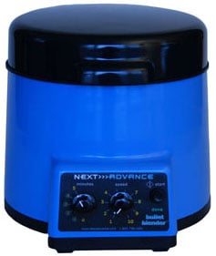
The Bullet Blender®
Save Time, Effort and Get Superior Results
- Consistent and High Yield Results
Run up to 24 samples at the same time under microprocessor-controlled conditions, ensuring experimental reproducibility and high yield. Process samples from 10mg or less up to 3.5g. - No Cross Contamination
No part of the Bullet Blender® ever touches the lymph node samples – the sample tubes are kept closed during homogenization. There are no probes to clean between samples. - Samples Stay Cool
Homogenizing causes only a few degrees of heating. Our Gold models keep samples at 4°C. - Easy and Convenient to Use
Just place beads and buffer along with your lymph node sample in standard tubes, load tubes directly in the Bullet Blender, select time and speed, and press start. - Risk Free Purchase
The Bullet Blender® comes with a 30 day money back guarantee and a 2 year warranty, with a 3 year warranty on the motor. The simple, reliable design enables the Bullet Blenders to sell for a fraction of the price of ultrasonic or other agitation based instruments, yet provides an easier, quicker technique.
Bullet Blender Homogenization Protocol for Lymph Node Tissue
Sample size |
See the Protocol |
| microcentrifuge tube model (up to 300 mg) | Small lymphatic tissue samples |
| 5mL tube model (100mg – 1g) | Medium lymphatic tissue samples |
Selected Publications for Lymphatic Tissue
See all of our Bullet Blender publications!
Hoover, C. E., Davenport, K. A., Henderson, D. M., Pulscher, L. A., Mathiason, C. K., Zabel, M. D., & Hoover, E. A. (2016). Detection and Quantification of CWD Prions in Fixed Paraffin Embedded Tissues by Real-Time Quaking-Induced Conversion. Scientific Reports, 6. https://doi.org/10.1038/srep25098
Falendysz, E. A., Lopera, J. G., Lorenzsonn, F., Salzer, J. S., Hutson, C. L., Doty, J., Gallardo-Romero, N., Carroll, D. S., Osorio, J. E., & Rocke, T. E. (2015). Further Assessment of Monkeypox Virus Infection in Gambian Pouched Rats (Cricetomys gambianus) Using In Vivo Bioluminescent Imaging. PLOS Neglected Tropical Diseases, 9(10), e0004130. https://doi.org/10.1371/journal.pntd.0004130
Liao, X., Ren, J., Wei, C.-H., Ross, A. C., Cecere, T. E., Jortner, B. S., Ahmed, S. A., & Luo, X. M. (2015). Paradoxical Effects of All-Trans-Retinoic Acid on Lupus-Like Disease in the MRL/lpr Mouse Model. PLOS ONE, 10(3), e0118176. https://doi.org/10.1371/journal.pone.0118176
Mitchell, D. A., Batich, K. A., Gunn, M. D., Huang, M.-N., Sanchez-Perez, L., Nair, S. K., Congdon, K. L., Reap, E. A., Archer, G. E., Desjardins, A., Friedman, A. H., Friedman, H. S., Herndon II, J. E., Coan, A., McLendon, R. E., Reardon, D. A., Vredenburgh, J. J., Bigner, D. D., & Sampson, J. H. (2015). Tetanus toxoid and CCL3 improve dendritic cell vaccines in mice and glioblastoma patients. Nature, 519(7543), 366–369. https://doi.org/10.1038/nature14320
Palomares, R. A., Hurley, D. J., Woolums, A. R., Parrish, J. E., & Brock, K. V. (2014). Analysis of mRNA expression for genes associated with regulatory T lymphocytes (CD25, FoxP3, CTLA4, and IDO) after experimental infection with bovine viral diarrhea virus of low or high virulence in beef calves. Comparative Immunology, Microbiology and Infectious Diseases, 37(5–6), 331–338. https://doi.org/10.1016/j.cimid.2014.10.001
Palomares, R. A., Parrish, J., Woolums, A. R., Brock, K. V., & Hurley, D. J. (2014). Expression of toll-like receptors and co-stimulatory molecules in lymphoid tissue during experimental infection of beef calves with bovine viral diarrhea virus of low and high virulence. Veterinary Research Communications, 38(4), 329–335. https://doi.org/10.1007/s11259-014-9613-2
Borschensky, C. M., & Reinacher, M. (2014). Mutations in the 3c and 7b genes of feline coronavirus in spontaneously affected FIP cats. Research in Veterinary Science, 97(2), 333–340. https://doi.org/10.1016/j.rvsc.2014.07.016
Goosen, W. J., Miller, M. A., Chegou, N. N., Cooper, D., Warren, R. M., van Helden, P. D., & Parsons, S. D. C. (2014). Agreement between assays of cell-mediated immunity utilizing Mycobacterium bovis-specific antigens for the diagnosis of tuberculosis in African buffaloes (Syncerus caffer). Veterinary Immunology and Immunopathology, 160(1–2), 133–138. https://doi.org/10.1016/j.vetimm.2014.03.015
Palomares, R. A., Brock, K. V., & Walz, P. H. (2014). Differential expression of pro-inflammatory and anti-inflammatory cytokines during experimental infection with low or high virulence bovine viral diarrhea virus in beef calves. Veterinary Immunology and Immunopathology, 157(3–4), 149–154. https://doi.org/10.1016/j.vetimm.2013.12.002
Melero, M., García-Párraga, D., Corpa, J., Ortega, J., Rubio-Guerri, C., Crespo, J., Rivera-Arroyo, B., & Sánchez-Vizcaíno, J. (2014). First molecular detection and characterization of herpesvirus and poxvirus in a Pacific walrus (Odobenus rosmarus divergens). BMC Veterinary Research, 10(1), 968. https://doi.org/10.1186/s12917-014-0308-2
Silver, A. C., Dunne, D. W., Zeiss, C. J., Bockenstedt, L. K., Radolf, J. D., Salazar, J. C., & Fikrig, E. (2013). MyD88 Deficiency Markedly Worsens Tissue Inflammation and Bacterial Clearance in Mice Infected with Treponema pallidum, the Agent of Syphilis. PLoS ONE, 8(8), e71388. https://doi.org/10.1371/journal.pone.0071388
Sahu, R. P., Petrache, I., Van Demark, M. J., Rashid, B. M., Ocana, J. A., Tang, Y., Yi, Q., Turner, M. J., Konger, R. L., & Travers, J. B. (2013). Cigarette Smoke Exposure Inhibits Contact Hypersensitivity via the Generation of Platelet-Activating Factor Agonists. The Journal of Immunology, 190(5), 2447–2454. https://doi.org/10.4049/jimmunol.1202699
Palomares, R. A., Walz, H. G., & Brock, K. V. (2013). Expression of type I interferon-induced antiviral state and pro-apoptosis markers during experimental infection with low or high virulence bovine viral diarrhea virus in beef calves. Virus Research, 173(2), 260–269. https://doi.org/10.1016/j.virusres.2013.02.010
Austin, W. R., Armijo, A. L., Campbell, D. O., Singh, A. S., Hsieh, T., Nathanson, D., Herschman, H. R., Phelps, M. E., Witte, O. N., Czernin, J., & Radu, C. G. (2012). Nucleoside salvage pathway kinases regulate hematopoiesis by linking nucleotide metabolism with replication stress. Journal of Experimental Medicine, 209(12), 2215–2228. https://doi.org/10.1084/jem.20121061
Bessen, R. A., Robinson, C. J., Seelig, D. M., Watschke, C. P., Lowe, D., Shearin, H., Martinka, S., & Babcock, A. M. (2011). Transmission of Chronic Wasting Disease Identifies a Prion Strain Causing Cachexia and Heart Infection in Hamsters. PLoS ONE, 6(12), e28026. https://doi.org/10.1371/journal.pone.0028026