Ideal for Skin Tissue Homogenization
Do you spend lots of time and effort homogenizing skin tissue samples? The Bullet Blender® tissue homogenizer delivers high quality and superior yields. No other homogenizer comes close to delivering the Bullet Blender’s winning combination of top-quality performance and budget-friendly affordability. See below for a skin tissue homogenization protocol.
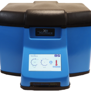
Save Time, Effort and Get Superior Results with
The Bullet Blender Homogenizer
Consistent and High Yield Results
Run up to 24 samples at the same time under microprocessor-controlled conditions, ensuring experimental reproducibility and high yield. Process samples from 10mg or less up to 3.5g.No Cross Contamination
No part of the Bullet Blender ever touches the tissue – the sample tubes are kept closed during homogenization. There are no probes to clean between samples.Samples Stay Cool
The Bullet Blenders’ innovative and elegant design provides convective cooling of the samples, so they do not heat up more than several degrees. In fact, our Gold+ models hold the sample temperature to about 4ºC.Easy and Convenient to Use
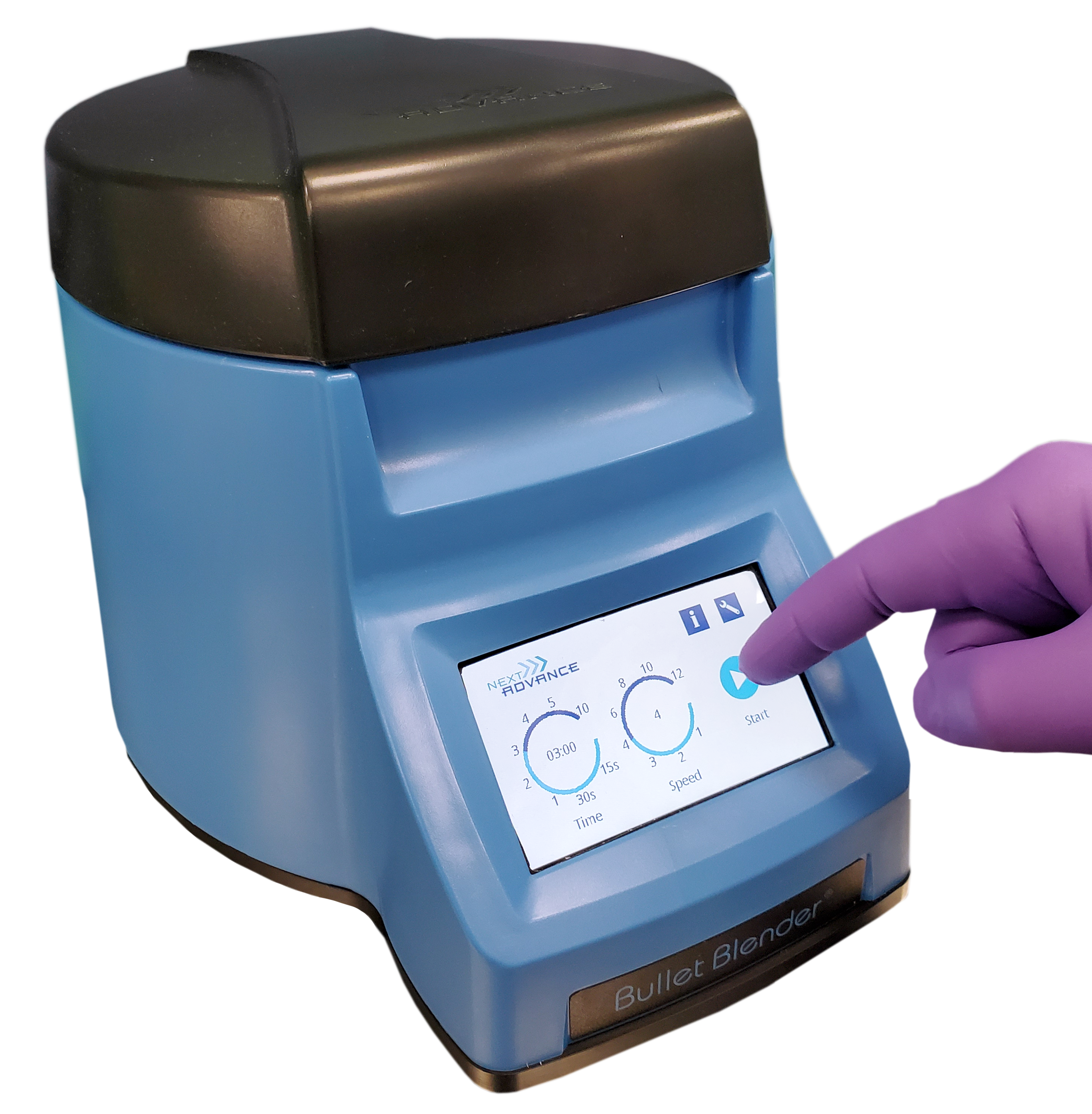
Just place beads and buffer along with your tissue sample in standard tubes, load tubes directly in the Bullet Blender, select time and speed, and press start.Risk Free Purchase
Thousands of peer-reviewed journal articles attest to the consistency and quality of the Bullet Blender homogenizer. We offer a 2 year warranty, extendable to 4 years, because our Bullet Blenders are reliable and last for many years.Skin Tissue Homogenization Protocol
| Sample Tube | Protocol |
|---|---|
| 1.5 mL tubes | 1.5 mL tubes Skin Protocol |
| 1.5/2 mL tubes using 5 mL adapters | 1.5/2 mL tubes using 5 mL adapters Skin Protocol |
| 5 mL tubes | 5 mL tubes Skin Protocol |
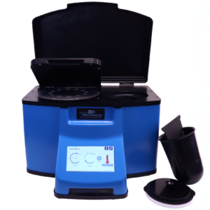
What Else Can You Homogenize? Tough or Soft, No Problem!
The Bullet Blender can process a wide range of samples including organ tissue, cell culture, plant tissue, and small organisms. You can homogenize samples as tough as mouse femur or for gentle applications such as tissue dissociation or organelle isolation.
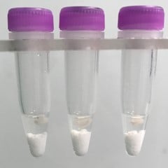
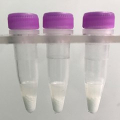
Skin tissue pieces (on beads in upper photo) are completely homogenized into the buffer (slightly darker in lower photo).
Want more guidance? Need a quote? Contact us:
Bullet Blender Models
Select Publications using the Bullet Blender to Homogenize Skin Tissue
Wen, L., Gao, Q., Ma, C., Ge, Y., You, L., Liu, R. H., Fu, X., & Liu, D. (2016). Effect of polysaccharides from Tremella fuciformis on UV-induced photoaging. Journal of Functional Foods, 20, 400–410. https://doi.org/10.1016/j.jff.2015.11.014
Kim, C.-H., Cheong, K. A., Lim, W. S., Park, H.-M., & Lee, A.-Y. (2015). Effects of low-dose light-emitting-diode therapy in combination with water bath for atopic dermatitis in NC/Nga mice. Photodermatology, Photoimmunology & Photomedicine, n/a-n/a. https://doi.org/10.1111/phpp.12220
Falendysz, E. A., Lopera, J. G., Lorenzsonn, F., Salzer, J. S., Hutson, C. L., Doty, J., Gallardo-Romero, N., Carroll, D. S., Osorio, J. E., & Rocke, T. E. (2015). Further Assessment of Monkeypox Virus Infection in Gambian Pouched Rats (Cricetomys gambianus) Using In Vivo Bioluminescent Imaging. PLOS Neglected Tropical Diseases, 9(10), e0004130. https://doi.org/10.1371/journal.pntd.0004130
Kuo, Y.-H., Chen, C.-W., Chu, Y., Lin, P., & Chiang, H.-M. (2015). In Vitro and In Vivo Studies on Protective Action of N-Phenethyl Caffeamide against Photodamage of Skin. PLOS ONE, 10(9), e0136777. https://doi.org/10.1371/journal.pone.0136777
Sebastian, R., Chau, E., Fillmore, P., Matthews, J., Price, L. A., Sidhaye, V., & Milner, S. M. (2015). Epidermal aquaporin-3 is increased in the cutaneous burn wound. Burns, 41(4), 843–847. https://doi.org/10.1016/j.burns.2014.10.033
Kim, C.-H., Kim, J.-Y., & Lee, A.-Y. (2015). Therapeutic and immunomodulatory effects of glucosamine in combination with low-dose cyclosporine A in a murine model of imiquimod-induced psoriasis. European Journal of Pharmacology, 756, 43–51. https://doi.org/10.1016/j.ejphar.2015.03.010
Danan-Gotthold, M., Golan-Gerstl, R., Eisenberg, E., Meir, K., Karni, R., & Levanon, E. Y. (2015). Identification of recurrent regulated alternative splicing events across human solid tumors. Nucleic Acids Research, 43(10), 5130–5144. https://doi.org/10.1093/nar/gkv210
Dao, V., Pandeswara, S., Liu, Y., Hurez, V., Dodds, S., Callaway, D., Liu, A., Hasty, P., Sharp, Z. D., & Curiel, T. J. (2015). Prevention of Carcinogen and Inflammation-Induced Dermal Cancer by Oral Rapamycin Includes Reducing Genetic Damage. Cancer Prevention Research, 8(5), 400–409. https://doi.org/10.1158/1940-6207.CAPR-14-0313-T
Mitchell, D. A., Batich, K. A., Gunn, M. D., Huang, M.-N., Sanchez-Perez, L., Nair, S. K., Congdon, K. L., Reap, E. A., Archer, G. E., Desjardins, A., Friedman, A. H., Friedman, H. S., Herndon II, J. E., Coan, A., McLendon, R. E., Reardon, D. A., Vredenburgh, J. J., Bigner, D. D., & Sampson, J. H. (2015). Tetanus toxoid and CCL3 improve dendritic cell vaccines in mice and glioblastoma patients. Nature, 519(7543), 366–369. https://doi.org/10.1038/nature14320
Besschetnova, T. Y., Ichimura, T., Katebi, N., St. Croix, B., Bonventre, J. V., & Olsen, B. R. (2015). Regulatory mechanisms of anthrax toxin receptor 1-dependent vascular and connective tissue homeostasis. Matrix Biology, 42, 56–73. https://doi.org/10.1016/j.matbio.2014.12.002
Krull, A. C., Shearer, J. K., Gorden, P. J., Cooper, V. L., Phillips, G. J., & Plummer, P. J. (2014). Deep Sequencing Analysis Reveals Temporal Microbiota Changes Associated with Development of Bovine Digital Dermatitis. Infection and Immunity, 82(8), 3359–3373. https://doi.org/10.1128/IAI.02077-14
Damodarasamy, M., Johnson, R. S., Bentov, I., MacCoss, M. J., Vernon, R. B., & Reed, M. J. (2014). Hyaluronan enhances wound repair and increases collagen III in aged dermal wounds: Hyaluronan and wound repair in aging. Wound Repair and Regeneration, 22(4), 521–526. https://doi.org/10.1111/wrr.12192
van der Plas-Duivesteijn, S. J., Mohammed, Y., Dalebout, H., Meijer, A., Botermans, A., Hoogendijk, J. L., Henneman, A. A., Deelder, A. M., Spaink, H. P., & Palmblad, M. (2014). Identifying Proteins in Zebrafish Embryos Using Spectral Libraries Generated from Dissected Adult Organs and Tissues. Journal of Proteome Research, 13(3), 1537–1544. https://doi.org/10.1021/pr4010585
Neely, C. J., Kartchner, L. B., Mendoza, A. E., Linz, B. M., Frelinger, J. A., Wolfgang, M. C., Maile, R., & Cairns, B. A. (2014). Flagellin Treatment Prevents Increased Susceptibility to Systemic Bacterial Infection after Injury by Inhibiting Anti-Inflammatory IL-10+ IL-12- Neutrophil Polarization. PLoS ONE, 9(1), e85623. https://doi.org/10.1371/journal.pone.0085623
Melero, M., García-Párraga, D., Corpa, J., Ortega, J., Rubio-Guerri, C., Crespo, J., Rivera-Arroyo, B., & Sánchez-Vizcaíno, J. (2014). First molecular detection and characterization of herpesvirus and poxvirus in a Pacific walrus (Odobenus rosmarus divergens). BMC Veterinary Research, 10(1), 968. https://doi.org/10.1186/s12917-014-0308-2
Dhall, S., Do, D. C., Garcia, M., Kim, J., Mirebrahim, S. H., Lyubovitsky, J., Lonardi, S., Nothnagel, E. A., Schiller, N., & Martins-Green, M. (2014). Generating and Reversing Chronic Wounds in Diabetic Mice by Manipulating Wound Redox Parameters. Journal of Diabetes Research, 2014, 1–18. https://doi.org/10.1155/2014/562625
Kim, C.-H., Cheong, K. A., & Lee, A.-Y. (2013). 850nm light-emitting-diode phototherapy plus low-dose tacrolimus (FK-506) as combination therapy in the treatment of dermatophagoides farinae-induced atopic dermatitis-like skin lesions in NC/Nga mice. Journal of Dermatological Science, 72(2), 142–148. https://doi.org/10.1016/j.jdermsci.2013.06.002
Silver, A. C., Dunne, D. W., Zeiss, C. J., Bockenstedt, L. K., Radolf, J. D., Salazar, J. C., & Fikrig, E. (2013). MyD88 Deficiency Markedly Worsens Tissue Inflammation and Bacterial Clearance in Mice Infected with Treponema pallidum, the Agent of Syphilis. PLoS ONE, 8(8), e71388. https://doi.org/10.1371/journal.pone.0071388
Kim, C.-H., Choi, Y.-S., Cheong, K. Ah., & Lee, A.-Y. (2013). Mechanism underlying the effect of combined therapy using glucosamine and low-dose cyclosporine A on the development of atopic dermatitis-like skin lesions in NC/Nga mice. International Immunopharmacology, 15(2), 424–432. https://doi.org/10.1016/j.intimp.2013.01.005
Song, J. S., Kim, S.-O., Kim, S.-H., Choi, H.-J., Son, H.-K., Jung, H.-S., Kim, C.-S., & Lee, J.-H. (2012). In Vitro and In Vivo Characteristics of Stem Cells Derived from the Periodontal Ligament of Human Deciduous and Permanent Teeth. Tissue Engineering Part A, 18(19–20), 2040–2051. https://doi.org/10.1089/ten.tea.2011.0318
Petreaca, M. L., Do, D., Dhall, S., McLelland, D., Serafino, A., Lyubovitsky, J., Schiller, N., & Martins-Green, M. M. (2012). Deletion of a tumor necrosis superfamily gene in mice leads to impaired healing that mimics chronic wounds in humans: LIGHT−/− mice wounds mimic human chronic ulcers. Wound Repair and Regeneration, 20(3), 353–366. https://doi.org/10.1111/j.1524-475X.2012.00785.x
von Grote, E. C., Venkatakrishnan, V., Duo, J., & Stenken, J. A. (2011). Long-term subcutaneous microdialysis sampling and qRT-PCR of MCP-1, IL-6 and IL-10 in freely-moving rats. Mol. BioSyst., 7(1), 150–161. https://doi.org/10.1039/C0MB00059K
Marji, J., O’Donoghue, S. I., McClintock, D., Satagopam, V. P., Schneider, R., Ratner, D., J. Worman, H., Gordon, L. B., & Djabali, K. (2010). Defective Lamin A-Rb Signaling in Hutchinson-Gilford Progeria Syndrome and Reversal by Farnesyltransferase Inhibition. PLoS ONE, 5(6), e11132. https://doi.org/10.1371/journal.pone.0011132
Nichols, D. P., Caceres, S., Caverly, L., Fratelli, C., Kim, S. H., Malcolm, K., Poch, K. R., Saavedra, M., Solomon, G., Taylor-Cousar, J., Moskowitz, S., & Nick, J. A. (2013). Effects of azithromycin in Pseudomonas aeruginosa burn wound infection. Journal of Surgical Research, 183(2), 767–776. https://doi.org/10.1016/j.jss.2013.02.003