Ideal for Tissue Dissociation
|
||
Save Time, Effort and Get Superior Results with |
||
 |
|
|
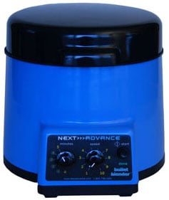 |
| Do you spend lots of time and effort homogenizing tissue samples? The Bullet Blender® is a multi-sample homogenizer that delivers superior results. No other homogenizer comes close to delivering the Bullet Blender’s winning combination of top-quality performance and budget-friendly affordability. | |
|
|
Bullet Blender settings for Tissue Dissociation
Sample size |
See the Protocol |
| microcentrifuge tube model (up to 300 mg) | Small spleen samples for generation of splenocytes |
Selected publications for Tissue Dissociation
See all of our Bullet Blender publications!
Booth, J. S., Salerno-Goncalves, R., Blanchard, T. G., Patil, S. A., Kader, H. A., Safta, A. M., Morningstar, L. M., Czinn, S. J., Greenwald, B. D., & Sztein, M. B. (2015). Mucosal-Associated Invariant T Cells in the Human Gastric Mucosa and Blood: Role in Helicobacter pylori Infection. Frontiers in Immunology, 6. https://doi.org/10.3389/fimmu.2015.00466
Booth, J. S., Toapanta, F. R., Salerno-Goncalves, R., Patil, S., Kader, H. A., Safta, A. M., Czinn, S. J., Greenwald, B. D., & Sztein, M. B. (2014). Characterization and Functional Properties of Gastric Tissue-Resident Memory T Cells from Children, Adults, and the Elderly. Frontiers in Immunology, 5. https://doi.org/10.3389/fimmu.2014.00294
Wiedner, S. D., Ansong, C., Webb-Robertson, B.-J., Pederson, L. M., Fortuin, S., Hofstad, B. A., Shukla, A. K., Panisko, E. A., Smith, R. D., & Wright, A. T. (2013). Disparate Proteome Responses of Pathogenic and Nonpathogenic Aspergilli to Human Serum Measured by Activity-Based Protein Profiling (ABPP). Molecular & Cellular Proteomics, 12(7), 1791–1805. https://doi.org/10.1074/mcp.M112.026534
Kim, K.-T., Zaikova, T., Hutchison, J. E., & Tanguay, R. L. (2013). Gold Nanoparticles Disrupt Zebrafish Eye Development and Pigmentation. Toxicological Sciences, 133(2), 275–288. https://doi.org/10.1093/toxsci/kft081
von Alvensleben, N., Stookey, K., Magnusson, M., & Heimann, K. (2013). Salinity Tolerance of Picochlorum atomus and the Use of Salinity for Contamination Control by the Freshwater Cyanobacterium Pseudanabaena limnetica. PLoS ONE, 8(5), e63569. https://doi.org/10.1371/journal.pone.0063569
Hong, N., De-Xing, Q., Deng-Xiang, Z., & Long-Zhong, R. (2012). A Simple Cultural Method for Detection of Mycoplasma bovis. Journal of Animal and Veterinary Advances, 11(10), 1643–1646. https://doi.org/10.3923/javaa.2012.1643.1646
Rayner, K. J., Suarez, Y., Davalos, A., Parathath, S., Fitzgerald, M. L., Tamehiro, N., Fisher, E. A., Moore, K. J., & Fernandez-Hernando, C. (2010). MiR-33 Contributes to the Regulation of Cholesterol Homeostasis. Science, 328(5985), 1570–1573. https://doi.org/10.1126/science.1189862