Ideal for Zebrafish Tissue Homogenization
Do you spend lots of time and effort homogenizing zebrafish tissue samples? The Bullet Blender® tissue homogenizer delivers high quality and superior yields. No other homogenizer comes close to delivering the Bullet Blender’s winning combination of top-quality performance and budget-friendly affordability. See below for a zebrafish tissue homogenization protocol.
Save Time, Effort and Get Superior Results with
The Bullet Blender Homogenizer
Consistent and High Yield Results
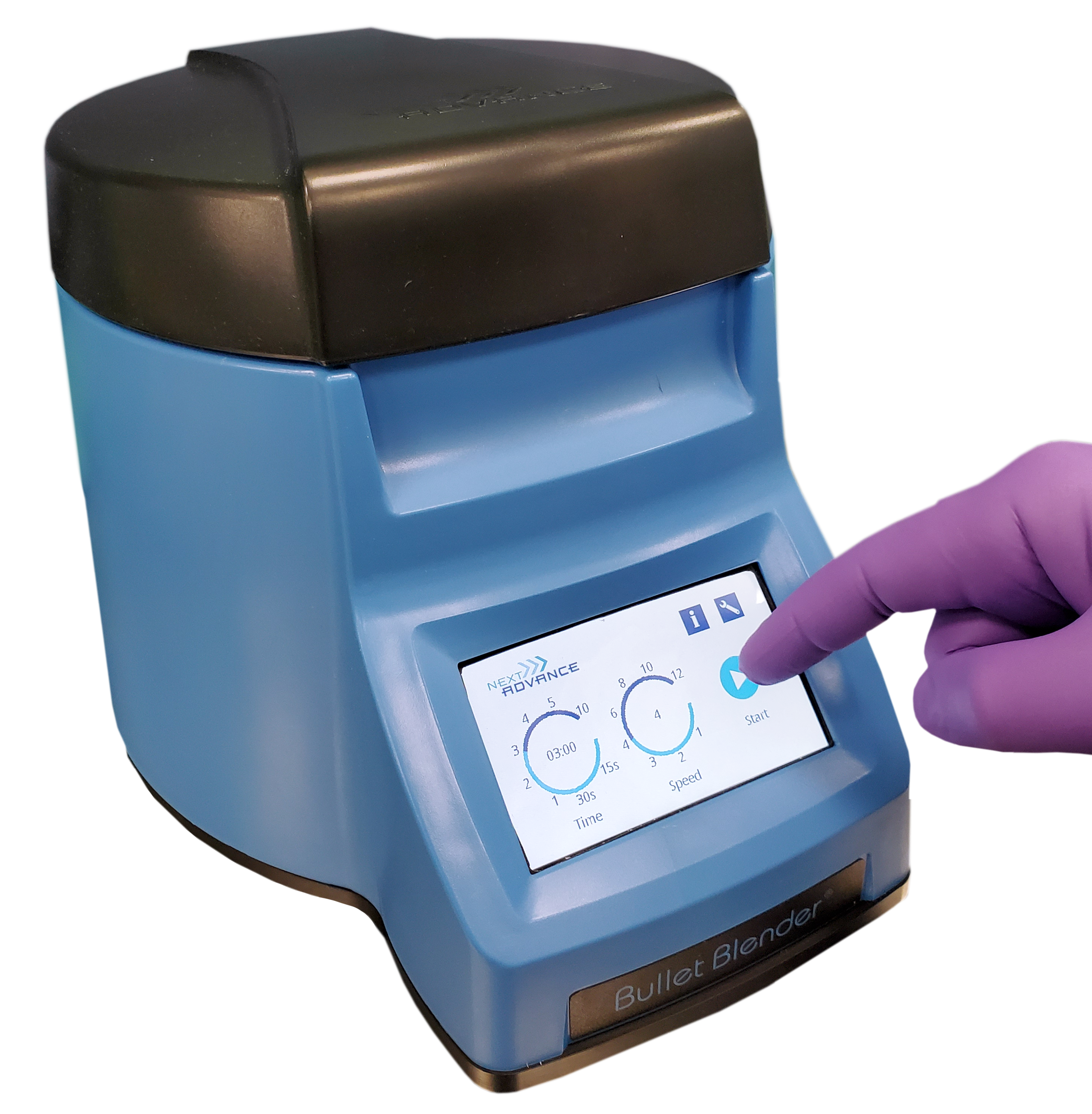
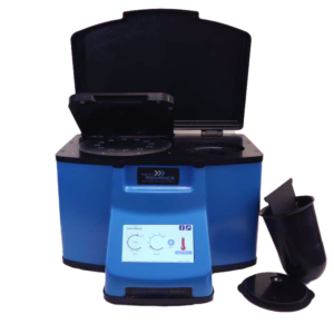
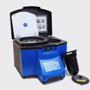
Run up to 24 samples at the same time under microprocessor-controlled conditions, ensuring experimental reproducibility and high yield. Process samples from 10mg or less up to 3.5g.No Cross Contamination
No part of the Bullet Blender ever touches the tissue – the sample tubes are kept closed during homogenization. There are no probes to clean between samples.Samples Stay Cool
The Bullet Blenders’ innovative and elegant design provides convective cooling of the samples, so they do not heat up more than several degrees. In fact, our Gold+ models hold the sample temperature to about 4ºC.Easy and Convenient to Use
Just place beads and buffer along with your tissue sample in standard tubes, load tubes directly in the Bullet Blender, select time and speed, and press start.Risk Free Purchase
Thousands of peer-reviewed journal articles attest to the consistency and quality of the Bullet Blender homogenizer. We offer a 2 year warranty, extendable to 4 years, because our Bullet Blenders are reliable and last for many years.Zebrafish Tissue Homogenization Protocol
Sample size |
See the Protocol |
| microcentrifuge tube model (up to 300 mg) | Small zebrafish/larva samples |
| 5mL tube model (100mg - 1g) | Medium zebrafish/larva samples |
What Else Can You Homogenize? Tough or Soft, No Problem!
The Bullet Blender can process a wide range of samples including organ tissue, cell culture, plant tissue, and small organisms. You can homogenize samples as tough as mouse femur or for gentle applications such as tissue dissociation or organelle isolation.
Want more guidance? Need a quote? Contact us:
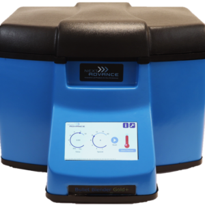
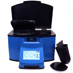
Bullet Blender Models
Select Publications using the Bullet Blender to Homogenize Zebrafish Tissue
Klucas, S. E., & Wong, R. Y. (2026). Differential Adenosine Signaling and Effects of Acute Caffeine Exposure on Alternative Stress Coping Styles in Zebrafish ( Danio rerio ). Brain and Behavior, 16(5), e71445. https://doi.org/10.1002/brb3.71445
Jiang, Z., El-Brolosy, M. A., Serobyan, V., Welker, J. M., Retzer, N., Dooley, C. M., Jakutis, G., Juan, T., Fukuda, N., Maischein, H.-M., Balciunas, D., & Stainier, D. Y. R. (2022). Parental mutations influence wild-type offspring via transcriptional adaptation. Science Advances, 8(47), eabj2029. https://doi.org/10.1126/sciadv.abj2029
Brinkmann, B. W., Beijk, W. F., Vlieg, R. C., Van Noort, S. J. T., Mejia, J., Colaux, J. L., Lucas, S., Lamers, G., Peijnenburg, W. J. G. M., & Vijver, M. G. (2021). Adsorption of titanium dioxide nanoparticles onto zebrafish eggs affects colonizing microbiota. Aquatic Toxicology, 232, 105744. https://doi.org/10.1016/j.aquatox.2021.105744
Robinson, C. D., Klein, H. S., Murphy, K. D., Parthasarathy, R., Guillemin, K., & Bohannan, B. J. M. (2018). Experimental bacterial adaptation to the zebrafish gut reveals a primary role for immigration. PLOS Biology, 16(12), e2006893. https://doi.org/10.1371/journal.pbio.2006893
Sarasamma, S., Audira, G., Juniardi, S., Sampurna, B., Lai, Y.-H., Hao, E., Chen, J.-R., & Hsiao, C.-D. (2018). Evaluation of the Effects of Carbon 60 Nanoparticle Exposure to Adult Zebrafish: A Behavioral and Biochemical Approach to Elucidate the Mechanism of Toxicity. International Journal of Molecular Sciences, 19(12), 3853. https://doi.org/10.3390/ijms19123853
McDougall, M. Q., Choi, J., Stevens, J. F., Truong, L., Tanguay, R. L., & Traber, M. G. (2016). Lipidomics and H218O labeling techniques reveal increased remodeling of DHA-containing membrane phospholipids associated with abnormal locomotor responses in α-tocopherol deficient zebrafish (danio rerio) embryos. Redox Biology, 8, 165–174. https://doi.org/10.1016/j.redox.2016.01.004
Chatzopoulou, A., Heijmans, J. P. M., Burgerhout, E., Oskam, N., Spaink, H. P., Meijer, A. H., & Schaaf, M. J. M. (2016). Glucocorticoid-induced attenuation of the inflammatory response in zebrafish. Endocrinology, en20152050. https://doi.org/10.1210/en.2015-2050
Techer, D., Milla, S., Fontaine, P., Viot, S., & Thomas, M. (2015). Influence of waterborne gallic and pelargonic acid exposures on biochemical and reproductive parameters in the zebrafish ( Danio rerio ): Influence Of Gallic and Pelargonic Acid Exposure. Environmental Toxicology, n/a-n/a. https://doi.org/10.1002/tox.22228
Doldur-Balli, F., Ozel, M. N., Gulsuner, S., Tekinay, A. B., Ozcelik, T., Konu, O., & Adams, M. M. (2015). Characterization of a novel zebrafish (Danio rerio) gene, wdr81, associated with cerebellar ataxia, mental retardation and dysequilibrium syndrome (CAMRQ). BMC Neuroscience, 16(1). https://doi.org/10.1186/s12868-015-0229-4
Wong, R. Y., & Godwin, J. (2015). Neurotranscriptome profiles of multiple zebrafish strains. Genomics Data, 5, 206–209. https://doi.org/10.1016/j.gdata.2015.06.004
Goodale, B. C., La Du, J., Tilton, S. C., Sullivan, C. M., Bisson, W. H., Waters, K. M., & Tanguay, R. L. (2015). Ligand-specific transcriptional mechanisms underlie aryl hydrocarbon receptor-mediated developmental toxicity of oxygenated PAHs. Toxicological Sciences, kfv139. https://doi.org/10.1093/toxsci/kfv139
Elie, M. R., Choi, J., Nkrumah-Elie, Y. M., Gonnerman, G. D., Stevens, J. F., & Tanguay, R. L. (2015). Metabolomic analysis to define and compare the effects of PAHs and oxygenated PAHs in developing zebrafish. Environmental Research, 140, 502–510. https://doi.org/10.1016/j.envres.2015.05.009
Peterman, E. M., Sullivan, C., Goody, M. F., Rodriguez-Nunez, I., Yoder, J. A., & Kim, C. H. (2015). Neutralization of Mitochondrial Superoxide by Superoxide Dismutase 2 Promotes Bacterial Clearance and Regulates Phagocyte Numbers in Zebrafish. Infection and Immunity, 83(1), 430–440. https://doi.org/10.1128/IAI.02245-14
Lindenburg, P. W., Ramautar, R., Jayo, R. G., Chen, D. D. Y., & Hankemeier, T. (2014). Capillary electrophoresis-mass spectrometry using a flow-through microvial interface for cationic metabolome analysis: CE and CEC. ELECTROPHORESIS, 35(9), 1308–1314. https://doi.org/10.1002/elps.201300357
van der Plas-Duivesteijn, S. J., Mohammed, Y., Dalebout, H., Meijer, A., Botermans, A., Hoogendijk, J. L., Henneman, A. A., Deelder, A. M., Spaink, H. P., & Palmblad, M. (2014). Identifying Proteins in Zebrafish Embryos Using Spectral Libraries Generated from Dissected Adult Organs and Tissues. Journal of Proteome Research, 13(3), 1537–1544. https://doi.org/10.1021/pr4010585
Tonyushkina, K. N., Shen, M.-C., Ortiz-Toro, T., & Karlstrom, R. O. (2014). Embryonic exposure to excess thyroid hormone causes thyrotrope cell death. Journal of Clinical Investigation, 124(1), 321–327. https://doi.org/10.1172/JCI70038
Goodale, B. C., Tilton, S. C., Corvi, M. M., Wilson, G. R., Janszen, D. B., Anderson, K. A., Waters, K. M., & Tanguay, R. L. (2013). Structurally distinct polycyclic aromatic hydrocarbons induce differential transcriptional responses in developing zebrafish. Toxicology and Applied Pharmacology, 272(3), 656–670. https://doi.org/10.1016/j.taap.2013.04.024
Knecht, A. L., Goodale, B. C., Truong, L., Simonich, M. T., Swanson, A. J., Matzke, M. M., Anderson, K. A., Waters, K. M., & Tanguay, R. L. (2013). Comparative developmental toxicity of environmentally relevant oxygenated PAHs. Toxicology and Applied Pharmacology, 271(2), 266–275. https://doi.org/10.1016/j.taap.2013.05.006
Kim, K.-T., Zaikova, T., Hutchison, J. E., & Tanguay, R. L. (2013). Gold Nanoparticles Disrupt Zebrafish Eye Development and Pigmentation. Toxicological Sciences, 133(2), 275–288. https://doi.org/10.1093/toxsci/kft081
Yang, J., & Xu, X. (2012). Actinin2 is required for the lateral alignment of Z discs and ventricular chamber enlargement during zebrafish cardiogenesis. The FASEB Journal, 26(10), 4230–4242. https://doi.org/10.1096/fj.12-207969
Saili, K. S., Corvi, M. M., Weber, D. N., Patel, A. U., Das, S. R., Przybyla, J., Anderson, K. A., & Tanguay, R. L. (2012). Neurodevelopmental low-dose bisphenol A exposure leads to early life-stage hyperactivity and learning deficits in adult zebrafish. Toxicology, 291(1–3), 83–92. https://doi.org/10.1016/j.tox.2011.11.001
Chen, C.-F., Chu, C.-Y., Chen, T.-H., Lee, S.-J., Shen, C.-N., & Hsiao, C.-D. (2011). Establishment of a Transgenic Zebrafish Line for Superficial Skin Ablation and Functional Validation of Apoptosis Modulators In Vivo. PLoS ONE, 6(5), e20654. https://doi.org/10.1371/journal.pone.0020654
Hoyt, C. (2009). Rapid, high-throughput homogenization of embryonic or larval zebrafish (Danio rerio). Protocol Exchange. https://doi.org/10.1038/nprot.2009.211
Techer, D., Milla, S., Fontaine, P., Viot, S., & Thomas, M. (2015). Acute toxicity and sublethal effects of gallic and pelargonic acids on the zebrafish Danio rerio. Environmental Science and Pollution Research, 22(7), 5020–5029. https://doi.org/10.1007/s11356-015-4098-2
Saili, K. S., Tilton, S. C., Waters, K. M., & Tanguay, R. L. (2013). Global gene expression analysis reveals pathway differences between teratogenic and non-teratogenic exposure concentrations of bisphenol A and 17β-estradiol in embryonic zebrafish. Reproductive Toxicology, 38, 89–101. https://doi.org/10.1016/j.reprotox.2013.03.009